Ultrasound technology has revolutionized modern livestock farming, providing a fast, non-invasive, and highly accurate method for monitoring animal health and reproduction. portable ultrasound machines, in particular, are a game-changer for farmers and veterinarians working in field conditions. Compact, battery-operated, and easy to transport, these devices help diagnose pregnancy, detect reproductive issues, and monitor internal organs in real time.

In this guide, we’ll walk through how to effectively use a portable ultrasound machine for various farm animals — including cattle, pigs, horses, sheep, and camels. Whether you're confirming pregnancy during the open period of a dairy cow or evaluating the health of a racehorse, mastering this tool can enhance your efficiency and herd health outcomes.
Getting Started: Understanding the Equipment
A portable ultrasound machine typically includes:
A probe or transducer
A monitor (either built-in or connected via cable/wireless)
A rechargeable battery pack
A gel for sound wave conduction
Straps or holsters for hands-free operation
Before use, ensure the machine is fully charged and the probe is clean and functioning. Select the appropriate probe frequency: lower frequencies (2.5–5 MHz) for deeper penetration in larger animals like cattle or camels, and higher frequencies (5–7.5 MHz) for small animals or superficial imaging.
Basic Operation Steps:
Apply ultrasound gel to the probe to eliminate air between the skin and the probe.
Restrain the animal safely to avoid movement and ensure accuracy.
Place the probe on the scanning site and gently adjust the angle for a clear image.
Interpret the image displayed in real-time on the monitor.

BXL-V50 Veterinary ultrasound Equipment
Now, let’s dive into species-specific guidelines.
Cattle (Beef and Dairy Cows)
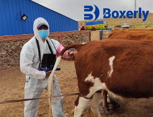
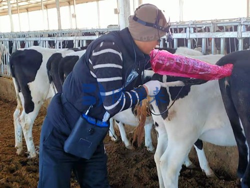
Ultrasound is commonly used in cattle for pregnancy diagnosis, especially during the “open” or “non-pregnant” period. Scanning can typically be done transrectally starting at 28–35 days post-breeding.

Steps:
Wear a long glove and lubricate your hand for rectal insertion.
insert the probe carefully into the rectum and locate the uterus through palpation.
Move the probe over the uterine horns to detect embryonic fluid or a fetus.
A clear black fluid pocket (anechoic) is a positive sign of early pregnancy.
Fetal heartbeat and limb movement can be seen after 35–45 days.
Benefits:
Reduces the open period by allowing early rebreeding.
Helps manage culling decisions and optimize herd fertility.
Pigs (Sows and Gilts)
In pigs, transabdominal scanning is usually done between 21–35 days post-mating. Use a convex or sector probe and scan the lower abdomen while the animal is standing.

Steps:
Apply gel on the lower side of the belly, just in front of the hind legs.
Position the probe at a 45-degree angle aiming toward the spine.
Look for multiple round fluid-filled sacs — these are embryonic vesicles.
Scan both sides of the abdomen to confirm pregnancy.
Key Notes:
Pigs can be scanned standing, with minimal stress.
Early detection allows better planning for farrowing space and feed management.
Horses (Mares)
Ultrasound in equine practice is used for ovulation monitoring, early pregnancy diagnosis, and reproductive tract evaluation.

Steps for pregnancy check:
insert the probe transrectally starting 14–16 days post-ovulation.
Look for a spherical fluid-filled vesicle in the uterus.
Confirm fetal heartbeat by day 25.
Repeat scanning at intervals to monitor fetal development and placental health.
Other Applications:
Monitor follicular development in breeding programs.
Detect uterine cysts, infections, or twin pregnancies.
Tips:
Keep the mare calm with proper restraint or mild sedation if necessary.
Use high-resolution probes for detailed ovarian assessment.
Sheep and Goats
Ultrasound is ideal for pregnancy detection in small ruminants starting around 30 days post-mating. A convex probe with a frequency of 5–7.5 MHz works well.

Steps:
Position the ewe or doe in standing or dorsal recumbency.
Apply gel on the lower abdomen near the udder.
Look for amniotic fluid pockets and embryo silhouettes.
Count the number of fetuses to plan nutrition and labor support accordingly.
Advantages:
Early pregnancy detection allows flock management by separating pregnant from open females.
Identifying multiples helps avoid underfeeding or overfeeding.
Camels
Camel reproduction management can be challenging due to longer gestation periods and less frequent estrus. Ultrasound is helpful in early pregnancy diagnosis (from around 18–20 days post-mating) and ovarian scanning.

Steps:
Use a rectal approach with a linear probe.
insert the probe gently and locate the uterine horns and ovaries.
A gestational sac and embryo can be visualized after 20–25 days.
Monitoring the dominant follicle helps time matings accurately.
Important Considerations:
Camels may require more restraint due to temperament.
Portable machines allow scanning in remote or desert locations with limited infrastructure.
General Tips for All Species
Always clean the probe before and after use with a disinfectant.
Label and save important images for record-keeping and veterinary review.
Monitor the battery level to avoid interruption during scanning.
Be patient and practice regularly to improve image interpretation skills.

Conclusion
Using a portable ultrasound machine effectively can significantly improve reproductive management, reduce costs, and enhance the productivity of your livestock operation. While species-specific techniques differ, the underlying principles of ultrasound use — proper positioning, patience, and anatomical knowledge — remain consistent. Investing time into learning this technology pays dividends in better herd health and farm profitability.
Whether you're checking a dairy cow during her open period, confirming pregnancy in a sow, or planning the breeding cycle of a mare, portable ultrasound machines put powerful insights directly in your hands.