In the world of equine sports and performance, few injuries are as feared—and as misunderstood—as a bowed tendon. Also known as a tendon strain or tendinitis, a bowed tendon typically refers to injury to the superficial digital flexor tendon (SDFT), which runs along the back of the cannon bone. It gets its name from the distinctive "bowed" shape of the leg when swelling occurs post-injury.

Fixing a bowed tendon on a horse is a multifaceted process involving accurate diagnosis, appropriate treatment, patient management, and long-term monitoring. The process is both art and science and is approached with slight variations in different parts of the world. In this article, we explore the causes, treatment strategies, and recovery protocols for bowed tendons, incorporating the international veterinary community’s best practices and insights. We’ll also highlight how advanced ultrasound technology like BXL’s DZ20 system plays a critical role in recovery.
Understanding the Injury: What Is a Bowed Tendon?
A bowed tendon occurs when tendon fibers—primarily those of the superficial digital flexor tendon—are overstretched or torn due to overexertion, fatigue, poor conformation, improper shoeing, or trauma. The injury is most common in racehorses, jumpers, and eventers, where repetitive high-speed or high-impact movements are common.
Clinical signs of a bowed tendon include:
Swelling in the mid-cannon region, often warm and painful to touch
Lameness of varying degrees
A visible “bow” or convex curve of the tendon
Limited range of motion in severe cases
Veterinarians in North America, Europe, and Australia all emphasize the importance of early recognition, as timely intervention significantly affects the prognosis.
Diagnosis: Ultrasonography Is Key
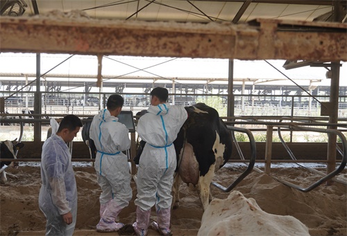
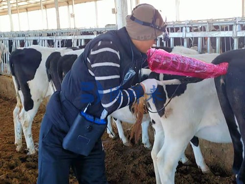
In almost every country, ultrasound imaging is considered the gold standard for diagnosing and grading the severity of bowed tendon injuries. While palpation gives clues, only ultrasound can reveal the internal fiber architecture, the extent of the tear, and involvement of surrounding tissues.
Veterinarians typically perform both longitudinal and transverse scans of the tendon, measuring:
Cross-sectional area (CSA)
Echogenicity (brightness)
Fiber alignment
Presence of core lesions or hemorrhage
An initial scan establishes a baseline, and follow-up scans help monitor healing. International experts agree: the more precise the imaging, the better the rehabilitation plan.

Treatment Phase 1: Acute Care (Days 1–14)
The first two weeks after injury are the most critical. The focus is on minimizing inflammation and preventing further damage.
Standard treatments include:
Stall rest with strict confinement
Cold therapy: Ice packs or cold hosing several times daily
Non-steroidal anti-inflammatory drugs (NSAIDs) such as phenylbutazone
Light bandaging to support the tendon and control swelling
In some countries like Germany and Japan, controlled magnetic therapy and pulsed electromagnetic field (PEMF) devices are also used during the acute phase to stimulate circulation and reduce edema.
Treatment Phase 2: Subacute and Regeneration Phase (Weeks 2–8)
During this stage, the inflammatory phase begins to subside, and fibroblasts begin to lay down new collagen fibers. The goal is to promote orderly tendon healing without creating excessive scar tissue.
Global practices may vary slightly, but common treatments include:
Controlled hand walking (5–10 minutes daily, gradually increased)
Therapeutic ultrasound or shockwave therapy
Laser therapy to stimulate tissue regeneration
Stem cell injections (more common in North America and the UK)
Platelet-rich plasma (PRP) therapy, used widely in Australia and the EU
Ultrasound monitoring every 2–3 weeks allows veterinarians to adjust exercise and therapy accordingly. A tendon healing poorly will show disorganized, hypoechoic (dark) areas with irregular fiber alignment. A tendon healing well will demonstrate narrowing lesion areas, improving echogenicity, and reestablishing linear fiber pattern.

Treatment Phase 3: Rehabilitation and Loading (Months 2–6+)
This is where recovery often falters—if pushed too hard or too soon, re-injury is likely. The global consensus among veterinarians is that tendon healing takes time, and that rest alone does not create strong, elastic scar tissue. Controlled exercise and progressive loading are essential.
Protocols usually involve:
Increasing walking duration to 30–40 minutes daily
Introducing trotting in straight lines around 4–5 months post-injury (if ultrasound supports it)
Frequent re-evaluation of the tendon via ultrasound
Gradual reintroduction to work by 6–8 months
Veterinarians in the U.S. often emphasize quantitative measurements and imaging, while practitioners in France or Italy may also stress the importance of visual symmetry, gait quality, and muscle development during rehab.

Surgical and Advanced Interventions
In some chronic or severe cases, surgical intervention may be necessary. This includes:
Tendon splitting to drain hemorrhage and promote neovascularization
Superior check ligament desmotomy to reduce strain on the SDFT
These procedures are more commonly performed in specialized clinics and usually accompanied by extensive rehabilitation. Regardless of geography, surgery is usually a last resort and is followed by rigorous ultrasound-guided monitoring.
Prognosis: What Can Be Expected?
The prognosis for a bowed tendon largely depends on:
Severity of the original injury
Quality and consistency of care
Timing of the intervention
The horse’s workload and discipline
Statistically, horses with mild to moderate lesions have a 60–80% chance of returning to previous levels of work if managed well. However, the recurrence rate is high—up to 30%—if the tendon is not given time to fully heal.

The Role of Ultrasound in Long-Term Recovery
Across all cultures and practices, one fact remains constant: ultrasonography is indispensable for both diagnosis and rehabilitation tracking. It is used to:
Determine when to increase workload
Detect early signs of reinjury
Evaluate tendon elasticity and fiber regrowth
Guide therapeutic decisions (e.g., timing of injections, suitability for return to sport)
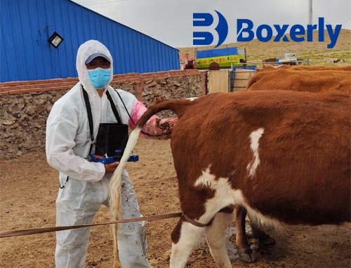
Portable, high-resolution systems such as the BXL-DZ20 allow practitioners to perform field scans during each stage of recovery. The BXL-DZ20’s clear imaging, portability, and tendon-specific presets make it an ideal solution for equine clinics, mobile vets, and rehabilitation centers. With real-time tendon imaging, veterinarians can confidently tailor each step of the recovery plan.
A Global Summary: Combining Patience with Precision
Fixing a bowed tendon on a horse is a long and complex journey. While treatment methods may vary slightly from country to country, the core principles—rest, inflammation control, controlled loading, and frequent imaging—are universally agreed upon. The key is a personalized, evidence-based approach that adapts to the individual horse’s healing process.
As equine medicine becomes increasingly sophisticated and globalized, technology plays a central role in improving outcomes. High-quality ultrasound imaging enables early diagnosis, informed decision-making, and safe return to work.

At BXL, we are proud to support the international veterinary community with cutting-edge tools like the BXL-DZ20 portable ultrasound system. Specifically designed for equine tendon and ligament evaluation, the BXL-DZ20 allows veterinarians to monitor recovery progress accurately, efficiently, and in real-time—no matter where in the world they’re working.
Whether you're treating a Thoroughbred in Kentucky, a dressage horse in Germany, or a show jumper in Dubai, BXL is your trusted partner in Veterinary ultrasound.