Ultrasound imaging has revolutionized veterinary medicine by providing a non-invasive, real-time window into the health of animals. Among the different types of ultrasound probes available, the linear probe stands out for its versatility and precision, especially when it comes to imaging superficial structures in a wide range of livestock, including cattle, pigs, horses, sheep, and even camels. This article explores the practical application of linear probe ultrasound in veterinary settings, focusing on its advantages, typical uses, and best practices for farm operations.
")
What is a Linear Probe Ultrasound?
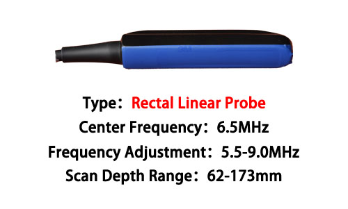
A linear ultrasound probe produces high-frequency sound waves (typically 5–15 MHz) that travel in a straight line, making it ideal for capturing high-resolution images of structures located near the surface of the body. Unlike convex or sector probes, which are better suited for deeper imaging, the linear probe is the tool of choice for detailed evaluations of tendons, muscles, joints, blood vessels, and superficial organs.
Key characteristics include:
High resolution for shallow depths (0–8 cm)
Flat contact surface for better skin contact
Broad field of view in a rectangular format
For livestock veterinarians and farm managers, understanding when and how to use a linear probe can significantly improve diagnostic accuracy and treatment outcomes.
Main Applications in Farm Animals
1. Musculoskeletal Imaging
In horses, injuries to tendons and ligaments are common, especially in performance animals like racehorses or working horses. The linear probe is indispensable for evaluating:
Superficial digital flexor tendons
Suspensory ligaments
Joint capsules and synovial structures
Similarly, in cattle and sheep, musculoskeletal injuries caused by rough terrain, breeding activities, or accidents can be effectively diagnosed with a linear probe.
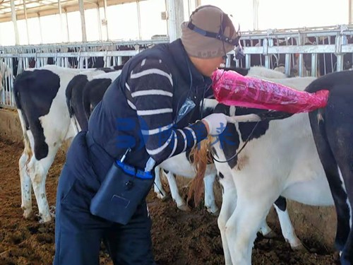
2. Reproductive Management
In pigs and sheep, reproductive efficiency is a top priority. Linear probes are excellent tools for:
Early pregnancy diagnosis (day 25–30 in pigs)
Follicle and corpus luteum assessment in ewes
Monitoring testicular size and structure in rams and boars
Accurate reproductive imaging helps farmers make better breeding decisions and improve overall herd productivity.
3. Soft Tissue and Superficial Organ Evaluation
For animals like camels, sheep, and cows, superficial abscesses, hematomas, and subcutaneous masses are not uncommon. The linear probe provides clear images for:
Measuring the size and internal characteristics of abscesses
Guiding needle aspiration or biopsy procedures
Evaluating superficial lymph nodes for signs of infection or neoplasia
In emergency situations, quick access to this kind of information can be life-saving.
4. Vascular Imaging and Blood Flow Assessment
Using Doppler mode in linear probes, veterinarians can assess blood flow in:
Jugular veins
Peripheral arteries
Umbilical vessels in newborn calves or foals
Vascular imaging is especially helpful for detecting thrombosis, monitoring perfusion, and evaluating critical care cases on the farm.
")
Advantages of Using a Linear Probe in Farm Settings
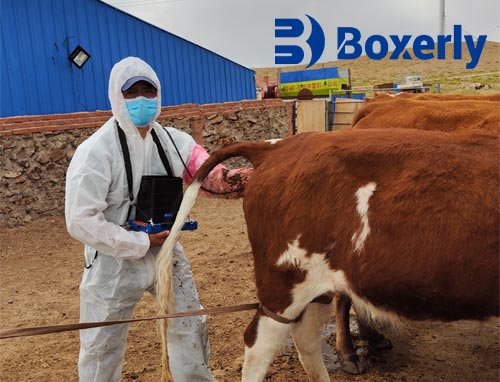
Portability: Devices like the BXL Veterinary ultrasound systems are compact, lightweight, and battery-operated, making them ideal for field use.
High-Resolution Imaging: Essential for detailed tendon, muscle, and reproductive evaluations.
Durability: Designed to withstand harsh farm environments — dust, humidity, and temperature changes are no match for modern veterinary ultrasounds.
Ease of Use: With minimal training, farm staff can learn to capture diagnostic-quality images, saving time and veterinary costs.
Best Practices for Effective Use
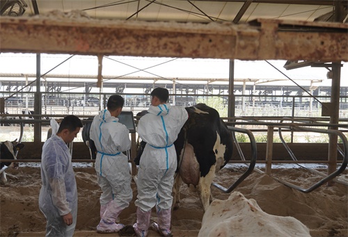
Proper Restraint: Always ensure the animal is securely and safely restrained to avoid movement during scanning.
Use Ample Ultrasound Gel: Good contact between the probe and the skin is critical for image quality.
Adjust Frequency Settings: For superficial imaging, set the probe to higher frequencies (10–15 MHz) to enhance resolution.
Scan in Multiple Planes: Always examine structures in longitudinal and transverse planes for a complete assessment.
Routine Cleaning: After each use, clean and disinfect the probe and ultrasound machine to prolong equipment life.
Future Outlook: Artificial Intelligence and Ultrasound in Veterinary Medicine
As technology evolves, ultrasound machines integrated with AI algorithms are becoming a game changer. Features such as automatic measurement of fetal parameters, tendon lesion recognition, and even early disease detection are already appearing in the latest BXL models. This means even faster and more accurate diagnoses at the point of care, right on the farm.
")
Conclusion
The linear probe ultrasound has become an indispensable tool in modern veterinary practice, particularly on farms where early diagnosis and treatment can mean the difference between success and failure. Whether it’s for monitoring a mare’s tendons, diagnosing a sow’s pregnancy, or checking a ram’s reproductive health, linear probes deliver precision, portability, and performance.
BXL veterinary ultrasound systems exemplify the best of this technology, providing reliable solutions that empower farm managers, livestock veterinarians, and rural communities to ensure the health and productivity of their animals.
Investing in the right ultrasound equipment today is an investment in the future of your herd.