There are many ways to detect whether a cow is pregnant or not. In the past, it was mainly by observing the characteristics of the pregnant cow, such as looking at the characteristics of the eyes, mouth, and tail. These are usually not accurate, and it takes two or three months for the cow to be bred. See it; some ask the veterinarian for rectal palpation. The accuracy and how many days after mating can be detected have a lot to do with the level and experience of the veterinarian.
Cattle farms will separate breeding cows from other cows for easy management. In addition, feed and various trace elements will be added to the bred cows so that the cows can get adequate nutrition during pregnancy. However, not every bred cow will be 100% pregnant, which causes a waste of feed. . If the non-pregnant cows can be detected early, the feeding supply of the non-pregnant cows can be reduced and the feeding cost can be saved. Therefore, if it is detected one day in advance, more feed can be saved. This may not be a big image for small farms. If there are hundreds of cattle farms, the daily feed cost is very high.
Therefore, many cattle farms now use veterinary B-ultrasound machines to detect the pregnancy of cows. The veterinary B-ultrasound machines have the advantages of less loss, short time, intuitive, convenient and accurate. It can accurately detect whether you are pregnant, and the imported machine can be shortened to about 27 or 28 days, mainly depending on the clarity of the machine.
Here’s how to use a veterinary ultrasound machine to detect whether a cow is pregnant or not:

- First, you need to do a good job of baoding, and it is better to tow the tested cow to the baoding rack.
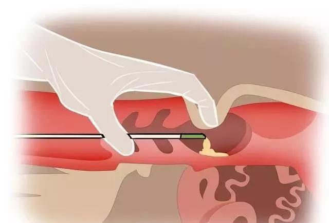
- Hold the Veterinary ultrasound probe in the palm of your hand in the direction of your fingers, with the index finger, middle finger, and ring finger above the probe. After entering the intestinal tube, gently press the probe to make it close to the intestinal wall. The thumb and little finger respectively pinch the two sides of the probe to fix and control the direction of the probe. Make sure that the underside of the probe is close to the intestine in the direction of ultrasound generation, and not blocked by fingers or feces. Clamp the probe with five fingers and rotate it through the anus and enter the rectum in a conical shape.
- After entering the rectum, slowly and gently move the probe back and forth to look for the bladder. After seeing the veterinary bladder on the monitor of the veterinary B-ultrasound instrument or wearing glasses, when the probe moves along the direction of the bladder to the position where the bladder gradually disappears, slowly Move the probe left and right to find the uterine wall. After finding the uterine wall, move the probe slightly to the left and right to observe the condition of the entire uterine wall while slowly advancing in the direction of the uterine wall. When it reaches the intestinal tube in the narrow part of the rectum (that is, the bifurcation of the uterine horn), the probe is tightly attached to the intestine and slowly rotates to the upper left direction. At this time, a circular cross section of the uterine horn can be observed, and continue to the direction of the uterine horn. On the upper left, you can observe and measure the left ovary, continue to rotate upward until the ovary disappears, and then return to the same way. When observing, the movements should be slow and gentle in order to observe the condition of the entire uterus and ovaries. After completing the left inspection, return to the bifurcation of the uterine horn in the direction of the left uterine horn, and rotate to the upper right to observe the condition of the right uterus and ovaries.
- When scanning the right uterine horn, the operation method is the same as that of the left. It is worth noting that the position of the right ovary is often slightly higher than that of the left, and it is more difficult to rotate the arm to the right. At this time, you must be patient and careful to complete the entire right side. Examination of uterine horns and ovaries.