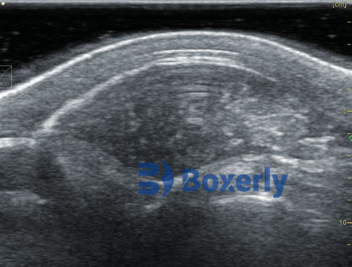
For the enhancement of contrast in animal B-ultrasound images, the dense grayscale distribution of the original image is made more dispersed, thereby increasing the contrast of the image and achieving a significant visual enhancement effect, making some details that were originally difficult to observe clear and distinguishable.
The contrast of animal B-ultrasound images is related to the distribution of bright and dark parts in the image. When most areas of an image are bright or dark (the latter is common in animal B-ultrasound), the animal B-ultrasound image shows a low contrast characteristic; When the proportion of brighter and darker parts in a Veterinary ultrasound image is comparable, the veterinary ultrasound image shows a high contrast characteristic.
Easy Scan veterinary ultrasound machine
Animal B-ultrasound images with low contrast are often composed of a limited range of gray levels, and their characteristic on the corresponding histogram is that the pixel value distribution is relatively concentrated and only occupies a small part of the available pixel range, resulting in a small dynamic range. When processing B-ultrasound images, first determine the range of the examination area, and then use the histogram corresponding to this area to determine which region the pixel values in the animal B-ultrasound image are mainly concentrated in. Use contrast stretching to correct the histogram of the original animal B-ultrasound image, so that the original image can utilize the entire grayscale range (i.e. 256 grayscale levels).
There are many methods for direct grayscale transformation, such as inverting veterinary ultrasound images, enhancing contrast, dynamic range compression, and grayscale segmentation. Enhancing the contrast of animal B-ultrasound images is actually to enhance the contrast of various parts of the original image. In practice, it is often achieved by increasing the dynamic range between two grayscale values in the original image.