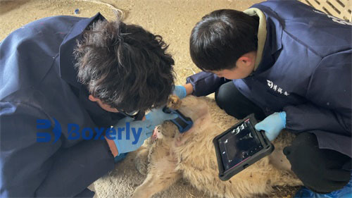
Ovarian cysts in ewes include two types: follicular cysts and luteal cysts. Both of these obstetric diseases can be diagnosed through sheep ultrasound. However, it is difficult to detect ovarian cysts in general sheep using ultrasound, and high-quality machines are needed to diagnose them, such as the BXL-V20 ultrasound machine used in sheep.
Cause: Follicular cyst is caused by degeneration of follicular epithelium, thickening of follicular wall connective tissue, death of oocytes, and delayed absorption of follicular fluid. Luteal cyst is caused by abnormal ovulation, leading to the luteinization of the follicular wall. Endocrine dysfunction, insufficient secretion of luteinizing hormone by the pituitary gland, and ovulation dysfunction in ewes can also lead to cysts.
Symptoms: Abnormal estrus in sheep with follicular cystic disease, short estrus cycle, prolonged estrus period, or persistent and intense estrus phenomenon. When sheep are examined by B-ultrasound, it can be found that there are large follicles on the cow's ovaries that exist for a long time. Sick sheep are restless, mooing, have decreased appetite, frequently excrete, chase or crawl over other ewes, and become infertile after mating. Upon dissection, it was observed that the ovaries had enlarged and contained 1-3 follicles with a wave like sensation. The sheep were diagnosed through B-ultrasound examination. The ewe with corpus luteum cyst showed no estrus, and dissection revealed an increase in ovarian volume and hardness upon touch.
Sheep B-ultrasound imaging
Treatment: Analyze the condition and treat based on syndrome differentiation. Treatment of follicular cysts: Formula 1, chorionic gonadotropin (HCG) 500-1000 international units per dose, intramuscular injection, combined with dexamethasone 8-10mg per dose. If one dose is ineffective, it can be repeated once. Formula 2, Ovulation Stimulating Hormone No. 3, 15-20 µ g per dose, intramuscular injection, once in the morning and once in the evening, for 3 consecutive days. Dexamethasone can be used in combination with each dose. After treatment, sheep ultrasound should be used to check the treatment status. Treatment of corpus luteum cyst: Formula one, 0.1-0.2mg of chloroquine per dose. Formula 2: Luteinizing hormone (LH) 50-100 international units per dose, intramuscular injection. If there is no improvement after one week of medication, the dosage should be increased, and after treatment, sheep ultrasound should be used to check the condition of the ovaries.