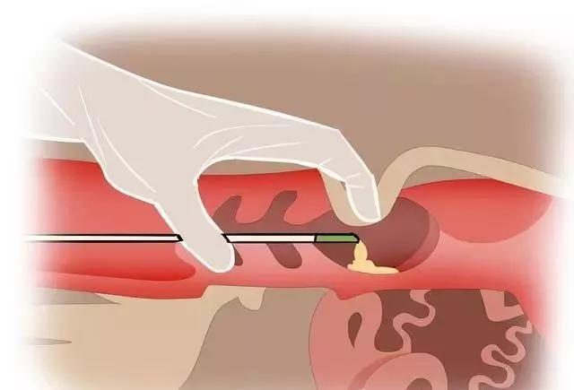
As an indispensable instrument for pig breeding, pig B-ultrasound machine is being used more and more widely. Then when you use it, do you have the following problems: 1. Can not find the location of the probe 2. The image will not look, will not analyze whether the pregnancy is 3, can not distinguish the number of days of pregnancy and a series of problems. In order to facilitate customers and solve users' problems, Boxiang animal B ultrasound Center shares the correct use of pig B ultrasound machine with graphics. The probe placement position of the pig B ultrasound machine: the probe is placed around the lower abdomen, the upper breast in front of the posterior flank, starting from the back of the last pair of mammary gland (commonly known as milk packet), with the improvement of pregnancy, the exploration site gradually moved forward, and finally up to the back end of the ribs. Scrape off dirt and dirt and apply coupler when probing. During rectal exploration, it is necessary to ensure that no coupling agent is required and the probe can be moistened with disinfectant water. That is, it points to the top of the last pair of teats of the sow (that is, the position of the uterus) 45 degrees to the front, 45 degrees to the side and the slope.
")
")
")
Pig B ultrasound machine sow image analysis:
1. Sow empty map features (as shown in Figure:) : Non-pregnant uterine horn is located at the left and right side of the bladder and below, and the bladder cavity is a non-echoic and regular liquid dark area. The wall of the non-pregnant uterine horn has weak ultrasound reflection, and its cross section ultrasonography shows various irregular circular weak reflection areas, but it is necessary to observe its boundaries to distinguish it from the cross section of the intestinal tube.
")
2. Image characteristics of the sow on the 18th day of pregnancy (as shown in Figure:) : 1 to 3 pregnancy sacs (pregnancy sac, liquid dark area) can be detected in the uterus of the sow on the 18th day of pregnancy, located in the front and bottom of the dark area of the bladder, the pregnancy sac is small, round or oval, the diameter of the dark area is less than 1 em, containing early fetal water, and the amount is small.
")
3. Images of sow pregnancy 20: on the 20th day of gestation, the sow's gestational sac is an oval liquid dark area with a diameter of 1-2 em and uneven edges
")
4. Image characteristics of sows on the 23rd day of pregnancy (as shown in Figure:) The pregnant sac in the uterus of sows on the 23rd day of pregnancy is a number of irregular round or oval dark areas with a diameter of 2 to 3 cm, the edges are not smooth, and some of them can detect embryonic spots, which are oval low-intensity echo light clusters or light spots
")
5. Image characteristics of the sow at 24 days of pregnancy (as shown in the figure:) Above the bright line is the uterine area. The above image shows three adjacent gestational sacs in the uterine area and also shows the fetal reflex.
")
6. On the 25th day of gestation, multiple irregularly shaped pregnancy sacs were detected, and two adjacent pregnancy sacs and their fetal reflex were shown in the uterine area.
")
")
7. Image characteristics of the sow at the 26th day of pregnancy (Figure:) Five adjacent pregnancy sacs are displayed simultaneously in the uterine area
")
8. Image characteristics of the sow at 28 days of gestation (Figure:) Four adjacent pregnancy sacs are displayed simultaneously in the uterine area. There are four pregnancy-sacs, the top of which is a large pregnancy-sac with significant fetal reflection images.
")
")
9. Day 32 gestation image: Two adjacent pregnancy sacs are shown simultaneously in the uterine area. Six gestational sacs are shown, with a large central gestational sac containing a strongly echoic fetal reflection image.
")
")
10. Not only can the pregnancy sac be detected on the 35d of pregnancy, but also the fetal body can be detected, with a diameter of 6 ~ 10cm. Fetal skeletal reflexes are enhanced. There is a flickering fetal movement, a sound shadow of enhanced bones.
")
11.45 day gestation image: There is a strong echo image of the fetal body in the right gestational sac in the uterine area. At this time, the bones have begun to calcify and the fetal tissues and organs have begun to differentiate. Visible foetus in the gestational sac
")
")
12.48 days of pregnancy image: After 45 days of pregnancy, the sow pregnancy test, amniotic fluid began to absorb and reduce, the piglets began to form, the image is complex and difficult to judge. At the same time, the average oestrus of the sow is 21 days, if the pregnancy is not determined within 21 to 42 days, it means that the pig farm has to raise another cycle to breed, resulting in great waste.
13. 55-day pregnancy image:
")
On day
14.65, a fetus's neck, chest and fetal heart beat were shown in the uterine area, which showed obvious differentiation of various tissues and organs, high degree of bone calcification, and obvious bone images.
")
")
Skeletal image of fetal head.
")
The whole section shows only the chest and abdominal cavity of a fetus, and the spine and thoracic cavity can be clearly shown, and the internal organs are obviously shown
")
Chest and rib imaging
15. 90-day gestation image
")
")
Cross sectional image with head up at 90 days old
")
16. 105-day gestation image
")
The entire section shows a skeletal image of the fetal thorax
")
Fetal heart section, clearly showing the heart section.