called the vaginal part of the cervix (or vagina), and the opening is called the external cervical os.
From the B-ultrasound image, the tissue structure of the uterus is divided into the serosa layer, muscle layer and mucosa layer from the outside to the inside. The serosa is the same as other gastrointestinal tracts. The muscles are mainly divided into two layers. The outer layer is longitudinal muscle fibers, and the inner layer is thicker and spiral circular muscle fibers. The cervical muscle is the attachment point of the uterine muscle and vaginal muscle, and it is also the sphincter of the uterus. Its inner layer is particularly thick and rich in dense collagen fibers and elastic fibers. The mucosa is composed of epithelium and lamina propria. The epithelium is a simple columnar cell (sheep also has pseudostratified columnar) cell. There are tubular, branched, and coiled uterine glands extending from the mucosal surface (except for the uterine caruncle) into the lamina propria. Its epithelium is a simple columnar cell with or without cilia. The motile cilia wave toward the vagina. Sheep have simple tubular glands. The secretory function of the gland is regulated by ovarian hormones. During estrus, estrogen increases the secretion of mucus and makes it thinner. During the period when the corpus luteum is in action, progesterone makes the mucus thicker. There are a large number of crypts on the cervical mucosa, where sperm survive for a long time. It is a storage depot for sperm, and there are also abundant nerve endings on the mucosa.
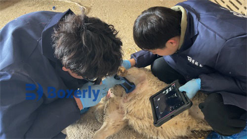
B-ultrasound images for sheep
The anatomical structure of the uterus varies not only with the changes in physiological functions, but also has many sheep species characteristics
For sheep with few parities, the uterine horns are curved like sheep horns and located in the pelvic cavity. After each delivery, B-ultrasound images show that the uterine horns do not completely return to their original shape and size, so the uterine horns of multiparous sheep are more or less stretched out and hang down into the abdominal cavity, with the front end of the horns pointed; the base diameter is 0.5~1.5 cm, and there is a longitudinal groove on the upper edge of the longitudinal septum between the bases of the two horns, called the inter-horn groove; the uterine body is short, only 3~5 cm long, and there are small hillock-like uterine caruncle on the mucosa of the uterine horns and uterine body, which is 80-100 in sheep, and there is often a shallow pit on its surface, so the maternal placenta formed by it is pelvic; the deep part contains rich blood vessels, and the cervical vaginal part protrudes into the vagina not long, only the upper and lower parts contain rich blood vessels, and the cervical vaginal part protrudes into the vagina not long, only the upper and lower parts protrude, and the upper part is larger. There are pigment spots composed of melanocytes on the mucosa of the uterine horns.
The cervix is well developed, 1-3 cm in diameter, with hard walls. The annular layer of the cervical muscle is very thick, and there is a dense network of blood vessels between it and the longitudinal layer. When it ruptures, it bleeds a lot. The mucosa and the annular muscle layer form 2-6 transverse crescent-shaped folds, which are wedged together to make the cervix spiral. The cervix contracts very tightly, and is more tightly closed during pregnancy. It only opens into a curved thin tube during estrus. There are also many low longitudinal folds on the mucosa, and the mucosa is divided into many folds by annular and radial grooves. The folds of multiparous sheep are sometimes enlarged like cauliflower.