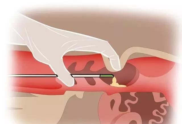
Sheep use B-ultrasound machine to observe the uterus. The uterus is the organ that nurtures embryos. During pregnancy, the uterine mucosa or a part of it forms the mother's placenta, which adapts to the needs of fetal development. The cervix is the opening of the uterus, which is tightly closed during pregnancy to protect the safety of the fetus. When the fetus develops and matures, the uterine muscles contract to expel the fetus from the body. In addition, the uterus also has functions such as transporting sperm and regulating luteal function.
The uterus consists of three parts: the horn, the body, and the neck. According to its different shapes, it can be divided into two types. Sheep use a B-ultrasound machine to observe that there is a mediastinum between the base of the two uterine horns of the sheep, and separating them is called a paired uterus. The uterine horn is curved, divided into major and minor bends. The uterine horn is 10-12 centimeters long, and the small curvature and the sides of the uterine body and neck are where the broad ligament of the uterus attaches, as well as where blood vessels and nerves enter and exit. The side opposite the lesser curvature is the greater curvature, and the anterior end of the uterine horn is connected to the fallopian tubes. The two corners converge backwards to form the uterine body. The anterior end of the cervix is connected to the uterine body, and its opening is called the cervical opening. The posterior end protrudes into the vagina, which is called the cervical vaginal part (or anal part), and the opening is called the cervical external opening.
Sheep use B-ultrasound machine to observe the tissue structure of the uterus, which is divided into serosal layer, muscular layer, and mucosal layer from the outside to the inside. Like other gastrointestinal tracts, the serosa is mainly divided into two layers: the outer layer is composed of longitudinal muscle fibers, and the inner layer is thicker, consisting of spiral circular muscle fibers. The cervical muscle is the attachment point of the uterine and vaginal muscles, as well as the sphincter muscle of the uterus. Its inner layer is particularly thick and rich in dense collagen and elastic fibers. The mucosa is composed of epithelium and particularly thick, dense collagen fibers and elastic fibers. The mucosa is composed of epithelium and lamina propria. The epithelium is composed of simple columnar cells (sheep also have pseudostratified columnar cells), with tubular, branched, and coiled uterine glands extending from the mucosal surface (except for the uterus) into the lamina propria. The epithelium is composed of simple columnar cells with or without cilia, with cilia fluctuating towards the vagina. Sheep have simple tubular glands. The secretion function of glands is regulated by ovarian hormones. During estrus, estrogen increases and dilutes the secretion of mucus. During the luteal phase, progesterone makes the mucus thick and sticky. There are a large number of crypts on the cervical mucosa, in which sperm survive for a long time and serve as a storage pool for sperm. There are also abundant nerve endings on the mucosa.
For sheep with fewer parity, the uterine horn is curved like a sheep horn and located in the pelvic cavity. After each delivery, the uterine horn does not fully return to its original shape and size, so the uterine horn of multiparous sheep extends to some extent and hangs down into the abdominal cavity, with a pointed and thin front end; The base diameter is 0.5-1.5 centimeters, and there is a longitudinal groove on the upper edge of the mediastinum between the two corner bases, called the intercornuate groove; The uterine body is short, only 3-5 centimeters long, and there are small mound shaped uterine masses on the uterine horns and mucous membranes of the uterine body. In sheep, there are 80-100 of them, and there is often a shallow pit on the surface, so the mother's placenta formed by it is bowl shaped; The deep part contains abundant blood vessels, and the cervix and vagina protrude into the vagina for a short period of time, with only 2 or 3 pieces protruding from the upper and lower parts, and the upper part being larger.
Sheep use B-ultrasound machine to observe pigment spots composed of melanocytes on the uterine horn mucosa. The cervix, which is 7 meters long and 7 meters long, is well-developed with a diameter of 1-3 centimeters and a hard wall. The circular layer of the cervical muscles is thick, and there is a dense vascular network between it and the longitudinal layer. When it ruptures, there is a lot of bleeding, and the mucosa and circular muscle layer form 2-6 horizontal crescent shaped folds that wedge together, making the cervix spiral. The cervix contracts tightly, closing even tighter during pregnancy and only opening as a curved thin tube during estrus. There are also many low longitudinal folds on the mucosa, which are divided into many folds by circular and radial grooves. The folds of sheep sometimes become enlarged like cauliflower