Examination of Follicle Atrophy in Cows by B-Ultrasound
Ovarian atrophy and alternate development in cows. Ovarian atrophy in cows refers to the cessation of follicular development halfway through. When observed by B-ultrasound, it can be seen that the diameter of the cow's follicles remains unchanged, and external estrous symptoms gradually disappear. Follicles alternate in development. In cows, it can be observed on ultrasound that follicles on one ovary of the cow stop developing, while there are varying numbers of follicles on the opposite or ipsilateral ovary that develop. However, before they mature, they begin to shrink, alternating one after another. In terms of treatment, 100-200 units of follicle stimulating hormone can be injected intramuscularly, once a day or once every other day, to promote follicular development, maturation, and ovulation. Alternatively, 5000-10000 units of chorionic gonadotropin can be injected intramuscularly, and the therapeutic effect can be observed through bovine ultrasound.
Endometritis in cows, with contaminated feces from the external genitalia and tail roots of lactating cows that have not been thoroughly cleaned and disinfected; When assisting with childbirth or removing the placenta, the surgeon's arms and instruments are not properly disinfected; Unclean delivery and breeding can cause postpartum endometrial infections, such as the breakdown of the placenta and stagnation of lochia. When observed by B-ultrasound in cows, strong echoes of inflammatory substances can be seen in the mucosal layer of the uterus. The best method for preventing and treating endometritis is postpartum injection of chloroquine to promote uterine smooth muscle contraction and lochia excretion, combined with the use of long-acting antibiotics to prevent postpartum infections. After treatment, the effect can also be observed by B-ultrasound in cows.
The ovaries of cows are in a quiescent state after being disrupted, with a size similar to fava beans and a normal or hard texture. There are no follicles or corpus luteum on the surface of the ovaries, and the uterus has weak contractions and reduced volume. The volume of the ovaries remains unchanged and no follicle formation can be observed using ultrasound in cows. For treatment, 100-200 units of follicle stimulating hormone can be injected intramuscularly, and 100-200 units of luteinizing hormone can be injected intramuscularly when estrus occurs.
Detection of follicles at different stages in cattle using B-ultrasound
The ovarian follicles of cows develop in the form of follicular waves, with dominant follicles maintaining their dominance for 4-5 days. On ultrasound, it can be observed in cows that the dominance is lost and degenerated during the estrus cycle of 11-12 days, lasting approximately 5-7 days. At the same time, the second wave occurs, selecting its dominant follicles and developing them to ovulation. However, in the three wave cycle, the second wave is replaced by the third wave, and ovulation occurs in the third dominant follicle. On B-ultrasound, it can be observed that dominant follicles maintain their morphological advantage (the largest follicle in both ovaries) for a longer period of time than maintaining their functional advantage (inhibiting the growth of other follicles).
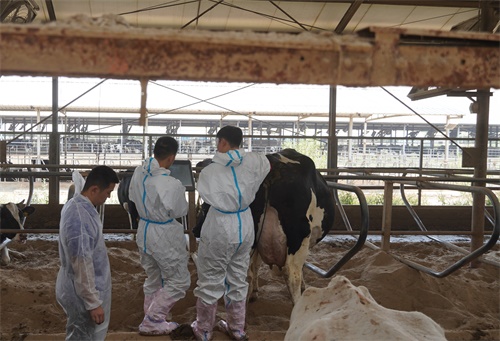
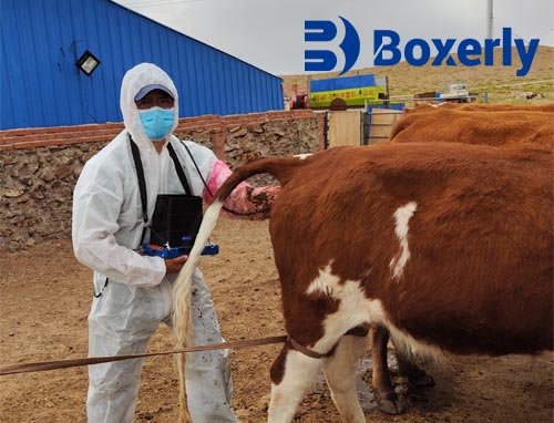
The cow follicle is a liquid structure on the ovary, which appears as a liquid dark area on the ultrasound image of cows and belongs to low-density echo. The follicular wall is composed of granulosa cell layers, and a normal follicular wall is relatively thin. Compared with traditional rectal examination, B-ultrasound in cattle presents significant advantages. Cattle can detect follicles with a diameter of less than 5mm on their ovaries using ultrasound. Continuous observation of ovarian activity can determine which stage of the estrus cycle they are in, thus determining the optimal time for estrus or insemination.
Before ovulation of animal follicles, bovine B-ultrasound examination can be used for timely monitoring to determine the appropriate time for insemination. During the estrus cycle, the dominant follicle diameter of cattle is generally 10-20mm, and ovulation occurs approximately 12 hours after estrus; During the estrus stage, the diameter of follicles varies from 14.2cm-15.9mm. When follicles develop to 21mm, cows begin to enter the ovulation stage; Sperm aspiration should be performed 6-24 hours after ovulation. Sheep and goat follicles are not as dominant as cattle. The diameter of the * * * large follicles in the luteal phase of goat follicle waves is smaller than that of the * * * large follicles after ovulation, and some may not even reach 5.0mm or more. However, they also have certain advantages in follicle function, and sheep and goats have certain similarities in follicle dominance. If mature follicles were present in the previous examination but disappeared in the second examination, ovulation occurs; Subsequently, there is corpus luteum development at the same point. To detect ovulation, it can be checked every 2 or 4 hours.