After a pig becomes pregnant, she will be pregnant for about three months, three weeks, and three days before giving birth to a litter of piglets. After the placenta of each embryo attaches to the uterus around day 21, embryonic cells begin to divide and differentiate into tissues and organs. Organs are formed at 35 days of embryonic development.
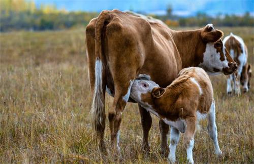
After weaning, sows can reproduce after feeding a litter of piglets. If she is not pregnant after * * * reproduction, you can reproduce her again when she is in heat again. The sow enters the second heating about 21 days after * * * heating. If she shows no signs of fever after three weeks, she is pregnant.
From the day after reproduction to the second day, the embryo migrates to the uterus to begin development. Pigs use ultrasound to observe embryos starting from single cells or fertilized eggs. Then divide until the embryo becomes a cell sphere. These cells differentiate into inner and outer cells. Outer cells become part of the outer membrane and placenta of the embryo, while inner cells develop into the fetus. At this stage, the cell sphere is called an embryo cell.
Between 12 and 20 days of pregnancy, the placenta develops and attaches to the uterus.
Do not feed large amounts of feed to sows during the first three weeks of pregnancy after reproduction, as this may reduce the chances of embryo survival.
Organ development: After placental attachment, pigs use B-ultrasound to observe that embryonic cells continue to divide and the embryo begins to grow.
Cells are divided into three layers:
The outer layers of the embryo are formed in external tissues, nervous system, and mammary gland cells.
In the mesoderm, it transforms into structural tissues, muscles, reproductive system, and these circulatory systems.
The embryo gradually transforms into internal organs, including the digestive system and liver.
The organ system of fetal pigs develops in a manner similar to that of human embryos. By the 35th day, the tissue had completed differentiation and formed organs.
Primary muscle fiber development continues until the 50th day. Pigs use B-ultrasound to observe the competition of embryos in the uterus for the necessary nutrients for muscle growth. Embryos without sufficient nutrition turned into tumors with less muscle mass than other piglets in the bedding.
The nutrition of sows can affect the development of embryos, especially the development of muscle fibers. After the first 21 days of pregnancy, increase the sow's feed according to her physical condition. When you can only feel firm pressure on your hips and spine, the sow can reach its ideal weight. After cell differentiation and organ formation, the embryo is usually considered a fetus.
***Post stage: Starting from the 90th day, pigs will quickly gain weight by observing the embryos with B-ultrasound. At this point, increase the sow's feed again so that she can maintain her weight and meet the nutritional needs of the fetus. At around 24 hours, the sow will give birth or self breed and become restless. She should have enough straw mats embedded. If you squeeze her nipples, there should be milk. Within one hour after delivery, blood may flow out of the sow's vagina.
Once * * * piglets are born, delivery should be completed within two to three hours, with one piglet born every 15 to 20 minutes. If there are signs of problems with the sow, such as prolonged periods between feeding piglets without the tension or fatigue experienced by piglets, she may need help. Although experienced operators can insert their hands into the delivery tube to help provide piglets, if your sow has problems, you should contact a veterinarian.