The attached are some ultrasound scans of umbilical abscesses in three month old calves of imported cattle using an ultrasound machine on grass, submitted by Dr. Paul Chard from Colorado (Colorado Brush) (published on Twitter @ CattleVet with Dr. Chard)
Dr. Paul Chard said, "The owner called me to take a look at this calf, and my biggest concern is that it may be a umbilical hernia. At that age, I rarely see umbilical abscesses on beef calves. Young calves are secondary to umbilical inflammation (postpartum umbilical cord infection). My most likely differential pre examination result is umbilical hernia
We tied this calf to the pasture with a rope using our head and heels. The tumor was too tight to effectively touch the contents of the abdomen, and there was a defect in the body wall. Within 10 seconds, I could make a clear diagnosis of umbilical cord abscess and puncture and discharge the abscess before the calf was compressed. I tried Aloka, "commented Dr. Chard.
")
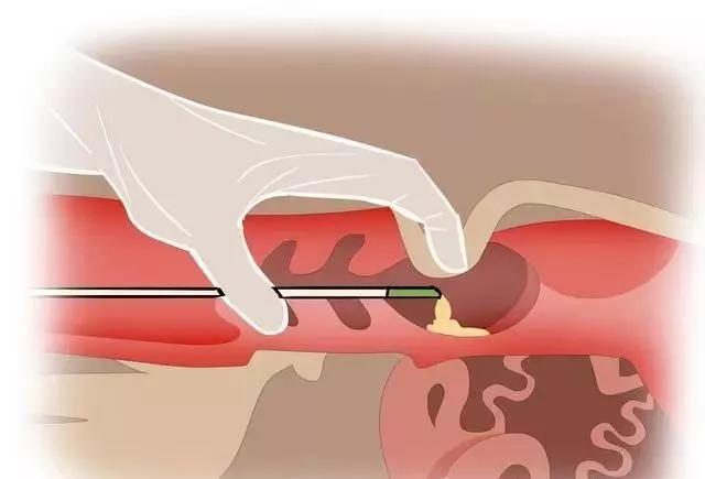
The image shows an abscess with a wall thickness of 7 millimeters and thickened skin. The displayed fluid is flocculent, indicating the presence of purulent fluid (pus). The umbilical cord is clearly visible, and if the abscess would be damaged by surgical drainage, then the umbilical cord is in good condition
In contrast, umbilical hernia can cause multiple intestinal peristalsis or
")
")
The omentum shown on the image is small
The Ibex portable ultrasound machine for imported cattle allows me to treat this animal more effectively and humanely in my own environment without leaving its mother to go to the clinic for examination.