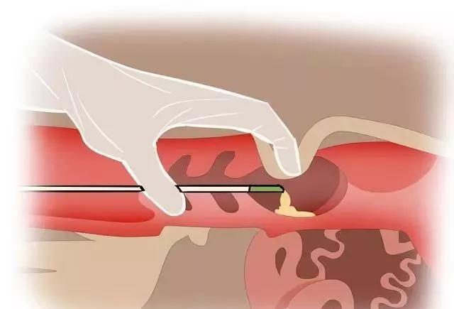
Veterinary B-ultrasound machine is one of the commonly used testing equipment in modern veterinary medicine, mainly used to detect tendons and pregnancy in horses. It can be accurately detected whether a mare is pregnant after 12 to 14 days of mating, while rectal palpation needs to be done after 35 days. It should be noted that rectal palpation of young mares can easily cause rectal tears, so rectal palpation is usually not a very safe choice. The mare prepares for transrectal ultrasound examination by wrapping her tail and emptying her rectum, then inserting the probe into the rectum and observing the cross-section of the uterus.
Veterinary B-ultrasound machine for pregnancy detection of mare
")
Animal B-ultrasound machine for detecting estrus in mare:
Because the eggs of a mare can only survive for about 12 hours, while the sperm can survive for up to 72 hours, it is wise to undergo ultrasound examination for ovulation. By using this technique, semen can be obtained within 12 to 24 hours before ovulation in the mare. The uterus of a mare, which is affected by estrogen during estrus, presents a typical "carriage wheel" appearance due to the swollen folds of the uterus. You can see the follicles on each ovary. Usually, follicles on one ovary become dominant. When its diameter reaches 35 to 40 millimeters, HCG or Ovuplant can be administered to the mare to induce ovulation. Then, after fertilization, the animal B-ultrasound machine can be used again to confirm ovulation.
In multiparous mares, rectal palpation remains important as it must be able to detect the consistency of developing follicles. In most cases, rectal palpation of small mares is not an option, so we rely solely on the size of ultrasound images.
Animal B-ultrasound machine for detecting pregnancy in mare:
Although the embryo can be seen from day 11 to 14, by day 15 to 17, the embryo vesicles have already been fixed on the uterine wall, making it easier to see. On the 24th day, the heartbeat can be seen to verify the presence of a live fetus. Early fetuses may be mistaken for uterine cysts, and vice versa. It is possible to regularly check whether the mare has increased in vesicle size. This can distinguish cysts from actual pregnancy. When realizing that the thermal cycle duration of a mare is about 21 days, the importance of ultrasound can be well understood.
Not being pregnant can decide to reproduce the mare in the subsequent estrus cycle. By the 60th to 78th day of pregnancy, fetal genital nodules (FGT) can be seen near the anus to confirm the presence of female foals, or male foals can be observed near the navel. Starting from the 80th day, transabdominal ultrasound can be used by placing the probe in the abdomen, just beside the head of the breast. Regular transrectal and transabdominal ultrasound examination of fetal heartbeat can be used to confirm a live fetus.
Although twins may be difficult to observe when they are placed side by side or overlapping in the uterus, we can usually imagine them as separate entities. Some data shows that 94% of twin pregnancies result in miscarriage or stillbirth of both fetuses. If twin pregnancies are allowed to occur without intervention, the mare usually brings the foal to eight or nine months, and then both fetuses will miscarry. Even if a mare brings twins to term, only 8% of people will be born and survive for more than two weeks.
If twin embryos of a mare are seen, they may be manually separated through the rectum and the smaller embryo may be pinched, leading to death. This program is usually done before the 17th day. Thoroughbred horses are notorious for having twins (3.5%), followed by standard horses at only 1%. Fortunately, small mares rarely have twins because it is difficult to reduce (pinch) the number of fetuses in this breed.
Veterinary B-ultrasound detection of pregnant mare ultrasound images
Animal B-ultrasound machine for detecting uterine cysts in mare:
Although uterine cysts are usually harmless, they can interfere with the attachment of the fetus to the uterus and can be easily observed with ultrasound. Intrauterine fluid retention caused by infectious or idiopathic endometritis can also be observed by transrectal ultrasound. The stimulation of some stallion semen on the mare's uterus is sufficient to cause endometritis. If fluid retention can be seen, the mare can be treated with intrauterine infusion, saline irrigation of the uterus, and/or oxytocin to increase the tension of the uterine body*** Afterwards, ultrasound examination can be used to evaluate the uterine tension of postpartum mares and their ability to 'reproduce' during subsequent 'pony fever'.