Fan shaped and linear arrays, as well as real-time B-mode ultrasound equipment, are commonly used for veterinary applications. Usually, linear probes are used together with linear imagers, while sector probes are used together with sector scanners. Scanners that can be used with sector and linear probes can also be used. A linear array probe has several piezoelectric crystals arranged in a row (which emit high-frequency sound waves when powered on), while a fan-shaped probe only has a few such crystals. Another major difference between the two systems is that the image generated by the linear array probe is rectangular on the screen, while the fan-shaped probe produces a pie shaped image corresponding to the scanning area. The commonly used method for scanning bovine reproductive organs is through the rectum, using a transrectal probe and a linear array scanner. For certain special applications, a fan-shaped probe is usually used for transvaginal scanning. Probes typically have frequency ranges of 3.0, 3.5, 5.0, and 7.5 MHz. The tissue penetration power and image resolution of sound waves depend on the frequency of the probe used. Therefore, a 3.0 MHz probe will provide greater tissue penetration but less detail, while a 7.5 MHz probe will provide smaller tissue penetration but greater resolution. The 5.0 MHz probe is a universal probe that can provide fairly detailed images of the ovaries and uterus.
Classification of probes for veterinary B-ultrasound machines
At present, the vast majority of veterinary B-ultrasound machines are easy to carry, but sturdy and durable scanners can withstand harsh on-site conditions, especially dust, moisture, sunlight, extreme temperatures, and voltage fluctuations, making them convenient for use on ranches. The weight of a typical animal ultrasound machine is around 1.5kg, and the built-in battery can be used continuously for 3-7 hours. Equipped with probes of 3.5, 5.0, and 7.5 MHz, choose the appropriate ultrasound probe according to the different detection animals and locations. Prices range from several thousand to tens of thousands, and even tens of thousands. When choosing, it is important to purchase a suitable veterinary ultrasound machine according to your own needs.
What can be detected by animal B-ultrasound machines?
Follicle development during animal estrus cycle
The growth and degeneration of ovarian follicles have long been a subject of speculation and debate, leading to conflicting hypotheses. Now, through the use of ultrasound, it has been determined that follicular growth occurs in a wavy pattern during the estrous cycle of cattle. Despite reports indicating the occurrence of two, three, and sometimes even four waves in each cycle, the two wave pattern in cows and the three wave pattern in heifers seem to be the norm. Follicle waves involve the synchronous growth of a group of follicles, where one follicle gains dominance over the other follicles and becomes the dominant follicle. Each dominant follicle has a growth phase and a quiescent phase, each lasting about 5-6 days*** The dominant follicle is anovulatory. It maintains its advantage for 4-5 days, usually losing its advantage and beginning to fade on the 1st or 12th day of the cycle. At the same time, the second wave of follicles has been recruited, and the selection of dominant follicles for the second wave has occurred. In the double wave cycle, this follicle continues to ovulate, while in the three wave cycle, the second dominant follicle also degenerates to make way for another group of follicles, known as the third wave.
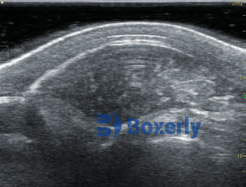
Ultrasound image of cow follicles
Ultrasound images of pregnancy in pigs, cows, and sheep
Animal B-ultrasound machines have been used to monitor and record the morphological changes of bovine fetuses at different stages of pregnancy. The study of bovine embryonic development relies on aborted fetuses or fetuses obtained during autopsy or slaughter. One of the early reports on the study of early embryonic development in cattle using real-time transrectal ultrasound examination. The length of embryonic vesicles gradually increases until day 26 when they begin to invade the contralateral horn. By the 32nd day, the embryonic vesicles had completely occupied the double horns. Heartbeat can be observed between the 26th and 29th day. During the period from day 20 to day 60, ultrasound recognizable features of the bovine pregnant body were detected, including the overall morphology of the fetus, fetal heartbeat, urinary bladder, spinal cord and umbilical cord, limb buds, amniotic membrane, optic vesicles, optic lens, fenders, ribs, and fetal movement.
Ultrasound images for detecting fetal age in cattle
Use transrectal ultrasound to describe changes in the size of fetal organs and body parts in cows, and record the relative frequency of different intrauterine positions between 30 days and 10 months of pregnancy. Until the fourth month, the frequency of anterior and posterior appearance is almost the same, with anterior appearance dominating during the period of 5-7 months. During the first four months of * * *, most of the major organs in the head, chest, abdomen, and pelvis can be seen through ultrasound. The accessibility of the last three body regions was later restricted. However, in approximately 80% of examination cases, the head can be touched at all stages of pregnancy. Liquid filled structures (eyes, skull, heart, and stomach) are easily recognizable because their contents have non echogenicity. Due to the limited field of view and penetration depth of sound waves, it is difficult to see the entire fetus in the later stages of pregnancy. For close visualization of the fetus, a 5 MHz probe was used, while a 3.5 MHz probe is more suitable for observing internal organs.
Animal ultrasound can detect male and female calves
One of the early reports that can visualize the male and female characteristics of bovine fetuses through real-time ultrasound. Between the 70th and 120th day of pregnancy in cows, the accuracy rate is 94%. Accurate male and female testing can be conducted after the 60th day. Testing males and females is suitable for singleton animals, as multiple births can affect accuracy.
Animal B-ultrasound machine for detecting male and female calves
Animal B-ultrasound machine can detect ovarian and uterine abnormalities
Follicular cysts display large (25-55 mm) non echogenic areas with very thin walls. On the contrary, corpus luteum cysts appear as hypoechoic areas surrounded by echogenic tissue of different thicknesses (2-5 millimeters). In cows with cysts treated with gonadotropin-releasing hormone, the thickness of the tumor wall increased from 2 millimeters to 6 millimeters within two weeks.
Bovine jaundice cyst
The animal B-ultrasound machine equipped with a dedicated OPU live egg collection probe can assist in guiding follicle aspiration work
This technology uses a 5.0 MHz sector probe. Cows with normal circulation and superovulation were used for transvaginal oocyte aspiration, achieving a recovery rate of 27.4% for oocytes. The ovaries of superovulated cows have been found to be easier to handle and aspirate, while puncturing small follicles is often difficult and risky, increasing the risk of rectal and even hand injuries for the cow operator.
Cattle live egg collection B-ultrasound machine