Fertilization of sows 12 to 0 hours before ovulation can achieve the best conception rate and litter size, so it is very important to use animal ultrasound machines to detect the development of cow ovaries.
Due to the reported differences in ovulation time among different facilities, management practices, genetics, and nutrition programs, it may be necessary to characterize this reproductive parameter for individual herds. Daily or twice estrus testing combined with ultrasound can promote the reproductive performance of females in livestock herds, as rapid identification of large follicles during estrus will indicate appropriate timing for fertilization.
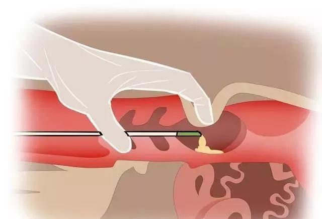
Animal B-ultrasound measurement of sows
Repeated ultrasound evaluation of the persistent presence of large follicles can assist producers in achieving an appropriate number of fertilizations. Due to reports that fertilization after ovulation may reduce reproductive performance, preventing reproduction after the disappearance of large follicles during estrus can improve overall productivity.
When using frozen sperm for artificial insemination, the fertilization time is crucial, and it is recommended to perform it within 8 hours before ovulation in sows to maintain an acceptable level of reproductive performance.
Only small (<3 mm) and/or medium-sized (3-6.5 mm) follicles were detected on the ovaries of reserve sows approaching puberty or weaned sows. In addition, identifying the ovaries of sows after weaning or even during estrus can help detect non cyclical sows with ovarian cysts and help eliminate them, as it has been reported that sows with multiple ovarian cysts are infertile. In the production environment, the ability to visualize the ovaries allows for the identification of ovarian cysts and even uterine infections, making it seem valuable for diagnosing reproductive failure. This technology may be very valuable, as cystic ovaries can account for 20% of reproductive failures in sows, while uterine abscess or acute uterine infection can account for 16% of reproductive failures.
Transrectal ultrasound machine detection is relatively easy to perform in many different types of production environments. Performing this technique in a standard pregnancy box is * * * practical. Understanding the reproductive status of breeding sows can help make reproductive management decisions regarding the timing and frequency of insemination, and prove useful for elimination decisions for sows and reserve sows. Experienced individuals can typically make decisions about ovarian and pregnancy status within 1 to 2 minutes. Valuable information can also be obtained from early pregnancy testing of pigs. For example, if it is found that females are not pregnant between the 18th and 21st day after mating, they can undergo stricter estrus testing and quickly reproduce during estrus. If they fail to express estrus, they can be eliminated. The ability to quickly determine pregnancy status through real-time imaging can also elucidate why animals that are determined to be pregnant between 21-25 days cannot maintain their pregnancy and subsequently return to estrus at irregular intervals. The ability to image the ovaries should help achieve higher accuracy in mating frequency and fertilization time, and subsequently improve delivery rates and litter size. Through informed, accurate diagnosis and proper management, * * * can ultimately reduce the non productive days of sows, improving the timing and frequency of elimination, gonadotropin treatment, and insemination.
The impact of veterinary B-ultrasound examination on obstetric practice is profound. Careful ultrasound examination can reveal important information about fetal anatomy, as well as fetal environment, growth, and health. Technology has evolved from generating two-dimensional images of the pregnant uterus to using Doppler methods to measure the blood circulation of the mother and fetus, and then to creating three-dimensional views of the anatomical structures of the mother and fetus.
")