One major advantage of using a cow ultrasound machine for detecting backfat eye muscle area is that it is non-invasive and relatively stress free for animals. Except for common equipment on farms or ranches and clinics, scanning operations do not require special handling equipment for animals. In order to alleviate animal stress and reduce the chances of injury to animals and operators, most people find that the best location for scanning operations is the livestock squeeze groove or squeeze area.
High definition backfat eye muscle area measuring device
Animals should be moved to the chute or compression area under as little pressure as possible. The head should be securely fixed in the head door and checked to ensure sufficient freedom and not cause suffocation or any other type of injury to the animal. After the operator safely immobilizes the animal, any previously described scanning operation can be performed, including backfat, hip fat, waist eye area, and muscle mass characteristics. Constrained beef cattle for scanning, scanning the area of the 12th and 13th ribs and buttocks.
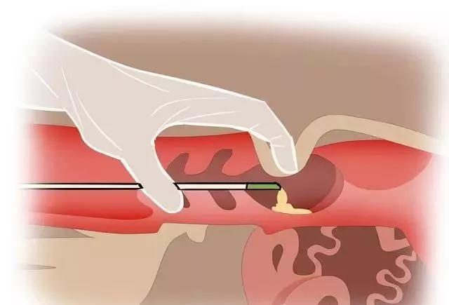
Measure the position of the backfat eye muscle in cattle
The site preparation for all scanning operations is similar. In order to achieve good acoustic contact, some operators like to cut off the hair on that area before scanning. Although this may be an ideal scenario for scanning purposes, it may not always be practical (i.e. time constraints, displaying or showcasing cattle, classifying cattle on the farm). If the scanned area has not been trimmed, the operator can improve image quality by using a brush or metal comb to ensure the removal of loose hair and dirt. Ultrasonic testing technicians/operators are very aware of the need for good acoustic contact between the probe and the skin surface, as sound waves do not propagate through the air. Human applications use scanning gel for acoustic contact, however, using gel for beef cattle scanning may be expensive and impractical because it is difficult to put gel into fur without air pockets. Therefore, scanning edible animals requires the use of vegetable oil (Figure 7). This coupling agent is easy to obtain, inexpensive, and harmless to animals, operators, or probes. (Do not use mineral oil as it is harmful to the probe surface and cable). Due to the fact that most scanning operations are performed under unfavorable outdoor environmental conditions, oil temperature may affect image quality. Clear results can be obtained when the oil temperature approaches 27 ° C. This can keep the oil flowing freely and better penetrate into the hair.
Ultrasound image of bovine eye muscle area
By observing the linear array probe, it is evident that some type of adaptation is necessary for cross-sectional scanning of the lumbar and ocular muscle areas. Therefore, acoustic contacts can be used as accessories for each unit. Contact points with different curvatures can be used for scanning beef or pigs. The contacts are easily attached to the probe and a small amount of scanning gel is used to ensure integrity. Application of palpation and acoustic coupling agent (vegetable oil) in scanning areas. The thermoelectric heating and cooling device suitable for maintaining scanning oil affects image quality, especially in cold climates. When the contact pad and acoustic gel are also kept near the oil temperature, a good effect can be obtained. After each use, warm soap and water cleaning pads should be used and stored in a suitable container to prevent damage and drying of flexible materials. If maintained and stored as recommended, the mat should be able to be used indefinitely.