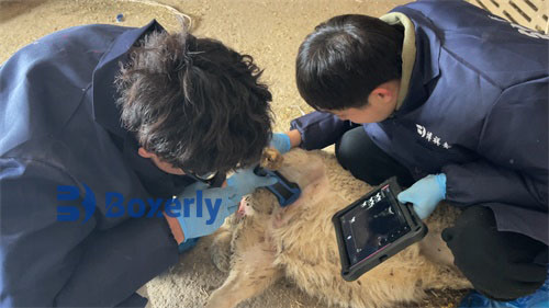
Prenatal condition: The cow is 3.5 years old and is a first-time cow with good nutritional status. The abdominal circumference is significantly enlarged before delivery, and the mammary glands are well developed with milk secretion. In the early stages, there were symptoms of restlessness, restlessness, and increased frequency and strength of restlessness, with a small amount of amniotic fluid flowing out. Later, the owner found that the frequency of restlessness decreased and the lips of the calf came out first. Oxytocin has been used, but it still cannot produce. Before processing, use bovine ultrasound to diagnose it.
Midwifery first involves vaginal examination or rectal examination using B-ultrasound for cows. The push-pull method may not be successful, but the baby's tongue can be felt with a little finger and there is a sensation of movement. The baby also has vital signs. If the midwifery fails, a cesarean section surgery will be performed.
Surgical procedure:
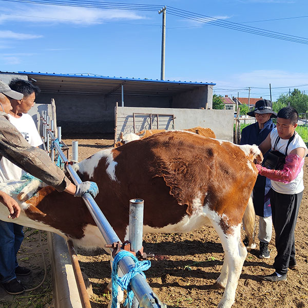
1. Surgical preparation: First, place the left side of the cow upside down in a stable position, shave a slightly larger area of the incision on the right thigh, rinse with clean water as much as possible, wrap the area outside the surgical area with sterile white cloth, and disinfect the shaved area with iodine.
2. The surgery is performed at the incision site (continuous between the bone width nodule and the navel), and 0.5% procaine 50ml is used for infiltration anesthesia. After about 5 minutes, a incision of about 30 centimeters is made, the skin is cut open, and the surgical handle is used to bluntly separate the thigh layers, fully stop bleeding, the peritoneum is cut open, the abdominal cavity is opened, and the peritoneum is pulled out of the incision and fixed. The abdominal cavity is examined, and the foot of the calf in the uterine horn is grasped by hand. It is pulled out as much as possible along the large bend of the uterine horn to the outside of the abdominal wall incision. A longitudinal incision of about 30 centimeters is made in the right uterine horn, avoiding the cotyledons, and the fetus is pulled out.
3. Peel off the placenta, complete the removal, dip the uterine slurry in sterile gauze, fill it with 800000 * 10 bottles of penicillin, then use intestinal thread to continuously suture the uterine serosa and muscle layer, and then use the inversion suture method to suture the second layer. After suturing, rinse the surface of the uterine horn with physiological saline and dry it, apply penicillin ointment, and send the uterus back to the abdominal cavity. Then use nodule suturing method to suture the peritoneum and various muscle layers, use tension reducing suturing method to suture the skin, and apply penicillin and fish fat ointment to the incision.