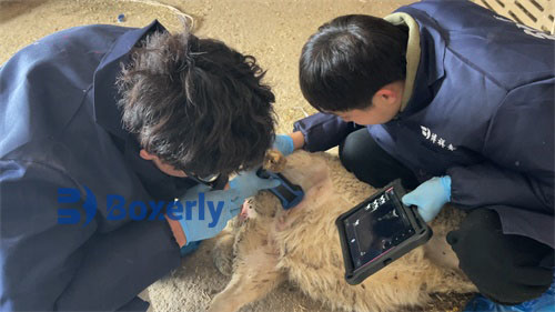
Standing or lying down on the sheep, the sheep's liver area detected by B-ultrasound on the right side of the body surface is sheared at the level of 8-10 intercostal shoulder joints, exposing the skin of the sheep's liver area detected by B-ultrasound. First, use a rechargeable electric hair clipper to remove coarse and long hair, then reduce the spacing density of the comb and cut off the remaining stubble. If necessary, apply an appropriate amount of coupling agent, then use a surgical knife to scrape the hair clean, and finally scan after applying the disinfectant.
Sheep should maintain sufficient and close contact with the skin using the B-ultrasound probe. Attention should be paid to avoiding human activity of the B-ultrasound probe and disturbance of the sheep. After scanning typical B-ultrasound images of sheep, freeze and save them, and record the scanning and storage time of the tomographic images. After freezing the B-ultrasound images of the sheep in a timely manner, relevant indicators were measured. The ultrasound images saved on the sheep ultrasound host can be transmitted to the computer via infrared through software for storage.
Calcified hepatic echinococcosis appears on the liver of sheep. The echo of the liver parenchyma is uneven, and the echo in the liver area of the sheep is detected as a light spot or spot by B-ultrasound scanning. The number of light spots varies with the course of the disease, and the volume, size, and shape are different. Echo enhancement appears in multiple places and multiple points, with a large contrast with the echo of normal liver tissue, showing partial or complete calcification
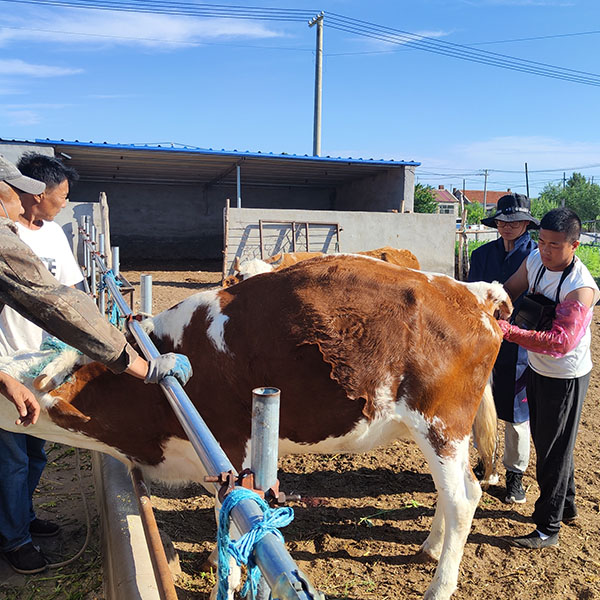
When operating sheep with B-ultrasound, we will find that it is relatively easy to use a portable charging shearing machine for rough wool sheep. Portable sheep ultrasound is easy to operate, highly efficient, and can easily detect clearer images. During scanning, the force is evenly applied and the liver area is scanned horizontally or vertically by sliding or rotating at a fixed point. During the process of using B-ultrasound to explore sheep, it was found that there were rib obstructions in the exploration area, especially when the sheep were thin and the ribs were clearly exposed, which affected the overall scan of the liver area. However, the sheep we used B-ultrasound to display were two-dimensional tomographic images, which resulted in a small portion of the fractures being difficult to display and may affect the diagnostic results.