People call high-frequency sound waves (2~10 MHz) that cannot be heard by the ear ultrasound, and created an imaging discipline that uses the physical properties of ultrasound for diagnosis and treatment, called ultrasound medicine. In the 1970s, my country applied this medical technology to the field of veterinary diagnosis. Veterinary ultrasound is divided into type A, type B, type D and type M, among which type B ultrasound examination is the most commonly used.
Principles and exploration methods of veterinary B-ultrasound
The principle of veterinary B-ultrasound is mainly to use the transducer (probe) to emit multiple beams of high-frequency ultrasound through the piezoelectric effect to penetrate into the cross-section of various tissues and organs for detection and generate echoes. The echoes can be received by the transducer and converted into high-frequency electrical signals and then transmitted to the host. After amplification, the cross-sectional image of the detected part is displayed on the screen, and there are light spots of different brightness on the image. Liquid absorbs sound waves, and the display screen is black. Dense tissue reflects most of the sound waves, and the display screen is white.
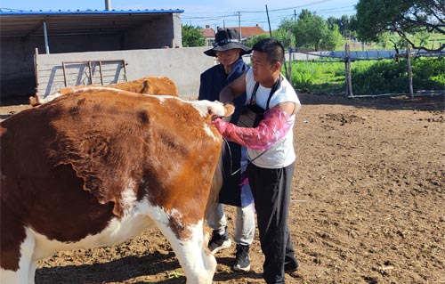
The application of veterinary B-ultrasound scanning in dairy cows includes in vitro exploration, vaginal exploration and rectal exploration. The most suitable method for dairy cow examination is rectal exploration. First, restrain the dairy cow to excrete the feces, and then lubricate the probe with lubricant. The operator stands directly behind the cow, opens the host and fixes it with his left hand, while holding the probe with his right hand and going deep into the rectum. First find the approximate location of the cow's reproductive organs, and then put the probe close to the top of the reproductive organs for exploration. During the inspection process, the ambient light should be moderate. If the light is too strong, cover the display screen with a cloth to produce an effective gray shadow effect.