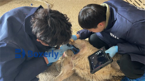
Imported backfat meters are widely used for detecting reproductive diseases and pregnancy in cows, but due to economic and technological reasons, the application of B-ultrasound to measure economic traits for beef cattle breeding and live evaluation is only carried out in a few research institutes and universities. At present, it is still in the stage of technological exploration and application summary. The following will summarize and exchange the key points and precautions of using ultrasound to detect backfat thickness and eye muscle area in pigs and live cattle.
Ultrasound waves usually refer to high-frequency vibration mechanical waves with a frequency higher than 20000Hz, and their propagation medium can be gas, liquid, or solid. Backfat instrument ultrasound has two forms: transverse wave and longitudinal wave. Longitudinal wave is a wave in which the vibration direction of the medium particles is parallel to the propagation direction of the wave, and it can cause changes in the volume of the medium.

High definition imported backfat analyzer
Because the ultrasound wave will produce reflection on the interface of different transmission media, the acoustic impedance of different media is different, the greater the difference of acoustic impedance, the stronger the ultrasound reflection, and the larger the light spot displayed; The more interfaces there are, the denser the light points displayed. By combining these light points, a cross-sectional acoustic image of the dielectric structure can be obtained.
The transmitting circuit in the imported backfat analyzer converts the current into pulse current through high-frequency oscillation and transmits it to the probe. The crystal inside the probe converts the pulse current into ultrasound waves through the "piezoelectric effect" and emits them; At the same time, the crystal inside the probe can also receive ultrasound waves reflected back from the interface of different media, and convert the ultrasound waves into electrical signals. After being processed by signal amplifiers and scanners, the sound image appears on the display (picture tube), which is called ultrasonic image.
There are various diagnostic methods for ultrasound image diagnosis, with quantitative diagnosis being the main application. The frequency of the probe can be divided into 3.5, 5.0, 7.5, and 10.0 MHz, with the more commonly used probe frequencies being 5.0 MHz and 7.5 MHz. The higher the frequency of the ultrasound probe of the backfat analyzer, the stronger its visibility and resolution; But the higher the frequency, the more significant the signal attenuation, resulting in shallower depths that ultrasound can transmit. When detecting deeper tissues or lesions, low-frequency probes should be used as much as possible, otherwise high-frequency probes should be used.
When using B-type ultrasound imaging diagnostic equipment, probes with frequencies of 3.5MHz and 5.0MHz are often used for body wall exploration. When conducting intracavitary exploration, probes with frequencies of 5.0MHz, 7.5MHz, and 10.0MHz are generally used, while also being as close as possible to the area being explored. The ultrasonic wave velocity of the imported backfat analyzer in various media is 1430m/s for fat, 1620m/s for muscle, an average of 1540m/s for soft tissue, and 3500rds for bones. The grayscale of the image displayed on the screen distinguishes the differences in tissues, with high-density tissues appearing white and medium density tissues appearing as gray pixels.
The selection principles for measuring backfat and eye muscles are: firstly, representativeness; Secondly, it is easy to detect in vivo; Thirdly, it is less affected by changes in animal position.