Application of animal B-ultrasound machine in dog breeding
")
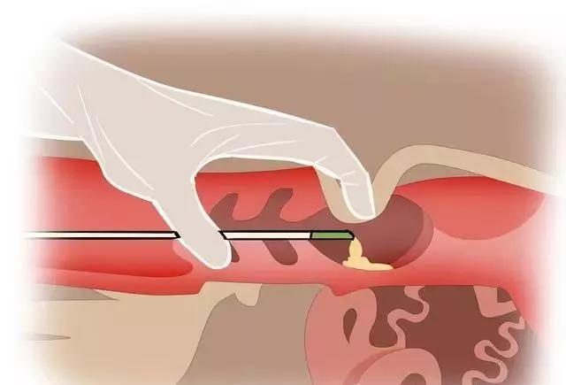
Animal B-ultrasound monitors ovarian development for timely mating, improving conception rate and increasing litter size. Dog ovaries are very small, generally 1.5 cm to 3 cm × 0.7 cm to 1.5 cm × 0.5 cm to 0.75 cm, and the outer capsule and fat are prone to echo interference. When exploring, it is necessary to find the correct position and observe carefully. The dog ovary is mulberry-shaped, located below the 3rd or 4th lumbar vertebra, 1 to 4 cm behind the kidney, and the position of the parous dog is further backward and downward. 2 to 3 days after estrus, ovaries containing developing follicles can be observed in the B-ultrasound section sonogram; the ovarian capsule with thin fat is clearly visible, the outer layer under the capsule has weak echo, and the central area has a pattern of strong echoes; the follicles are multiple circular echo-free areas on the periphery, with a maximum diameter of up to 13 mm. In the first and second stages of estrus (about 8 days after estrus), the follicles of dogs grow slowly. In the third stage (about 10-15 days after estrus), the follicles grow rapidly and then stop growing. At this time, ovulation is about to occur, which is the best time for mating or artificial insemination. When B-ultrasound is used to monitor ovarian development, the number of mature follicles (diameter is more than 3 mm, protruding from the capsule) can be calculated from multiple sections. When there are too few mature follicles (less than 5) or the number of synchronously growing follicles is small and the speed is slow, gonadotropins (such as FSH or PMSGLH or HCG) can be used in moderation to promote ovarian development and ovulation, improve conception rate, and increase litter size. Animal B-ultrasound is used to observe embryonic development, thereby estimating the number of fetuses, predicting fetal age, identifying stillbirths, and monitoring delivery. B-ultrasound can be used to observe the external structure of the embryo (such as uterus, gestational sac, placenta, fetal membranes and umbilical cord), the appearance of the embryo (such as fetal outline, limbs, external genitalia and fetal movement), and the internal structure of the embryo (such as fetal heartbeat, internal organs and bones). The gestational sac can be seen in dogs 15-20 days after pregnancy, the embryo can be seen in 23-25 days (a bright mass with strong echoes appears in the chorionic sac), the fetal heartbeat can be observed in 21-28 days, the head and trunk can be distinguished in 26-28 days, the limbs and the choroid plexus can be distinguished in 31-35 days, ossification begins in 35-40 days, and the optic vesicle appears and the internal organs can be distinguished in 40-47 days. The number of fetuses can be estimated based on the number of GS and embryos around 23 days; the gestational age can be predicted based on the echo structure that can be identified in the B-ultrasound section; the fetal heartbeat and embryo structure can be used to distinguish dead fetuses, emphysema fetuses, and embryo absorption and abortion. The internal structure of the uterus during and after delivery can be used to monitor delivery, determine whether delivery is over, and monitor the postpartum involution of the uterus.
Animal B-ultrasound diagnosis of reproductive diseases. There are many reproductive diseases in dogs, the most common of which are uterine pyometra and hydrops, ovarian cysts, dystocia and abortion, uterine, breast, testicular tumors, and prostate hypertrophy and tumors. B-ultrasound can be used to make a diagnosis or differential diagnosis, and the location, size, and characteristics of the diseased tissue can be qualitatively and quantitatively analyzed.
Veterinary B-ultrasound machine detection of canine diseases
Canine diseases are numerous and complex. Not all diseases can be diagnosed with imported B-ultrasound EVO. Here are several types of diseases with distinctive significance for B-ultrasound diagnosis.
Animal B-ultrasound machine detection of space-occupying lesions in and on the body of dogs
Space-occupying lesions refer to the growth and accumulation of diseased tissues that do not exist under normal circumstances between normal tissues, occupying the original position of normal tissues, thereby affecting the normal development and function of the body. Such as tumors, hematomas, abscesses, cysts, brain hydatids, etc., they can occur on the surface of the body, in deep tissues and internal organs, but no matter where they occur, their structure is always very different from normal tissues. B-ultrasound can just discover their existence, location, size and even their nature through the cross-sectional images, playing the role of a "scout", thus providing accurate and effective information for their overall elimination.
Fluid lesions in the chest, abdomen, pericardium, ventricles and luminal organs
One major feature of B-ultrasound is that when ultrasound encounters liquid tissue during transmission, it is almost completely absorbed or penetrated, and no reflected echo is generated, thus presenting a liquid dark area on the B-ultrasound cross-sectional sonogram. When fluid accumulates in places where fluid should not accumulate under normal circumstances, B-ultrasound accurately shows it and can show the degree of fluid accumulation and the formed elements mixed in the fluid. Such as pleural effusion, ascites; pericardial effusion, lateral ventricle hydrops; renal pelvic urine, bile duct fluid dilatation, pathological accumulation of gallbladder or bladder, upper intestinal effusion during intussusception obstruction, uterine pyometra, etc.
Animal B-ultrasound machine detects canine bile duct, urinary tract and gastrointestinal calculi
Cholecystolithiasis, bile duct calculi, kidney calculi, bladder calculi, urethral calculi, intestinal calculi, etc. are mostly caused by the deposition of minerals on the core material, condensing into relatively dense stone masses or mud sand, which forms an ultrasonic interface with a great impedance difference with the surrounding tissues or liquids, forming a light mass or light spot with a very strong echo in the B-ultrasound section sonogram, and often with an acoustic shadow. It is not difficult to confirm as long as the location is accurate during exploration. Strong echo light mass, acoustic shadow, and possible displacement of body position change are the three major characteristics of diagnosing stone disease.
Animal B-ultrasound machine detects substantial lesions of canine solid organs, tissues and internal organs
When solid organs such as liver, spleen, muscle, lymph node, testicles, etc. are hardened, calcified, softened, or liquefied, the B-ultrasound echo of the lesion site is either enhanced or weakened compared with the adjacent healthy tissue or the same organ on the opposite side. It is called substantial echo change in ultrasound diagnosis.
Animal B-ultrasound machine detects canine heartworm disease and cardiovascular disease in dogs
Canine heartworm disease is widely distributed in my country. It is caused by canine heartworms parasitizing in the right ventricle and pulmonary artery. Defects of the atrioventricular septum, atrial septum, and ventricular septum, stenosis of the tricuspid valve and mitral valve, and insufficiency are often encountered in clinical practice and are difficult to diagnose. M-mode echocardiography and ultrasound Doppler detection technology are one of the fast and effective means. Most B-ultrasound machines have the function of M-ultrasound and can also make a diagnosis.
Animal B-ultrasound machine detects gastrointestinal foreign bodies and damaged foreign bodies in dogs
Dogs are omnivorous animals and are active and playful. It is not uncommon for them to accidentally swallow children's toys, glass balls, hair balls, nylon ropes, tapes, etc., which may cause gastrointestinal foreign bodies or even obstructions. It is also common for dogs to eat bones, carrots, etc. and have esophageal obstructions. It is very necessary to judge the size, location, and properties of foreign bodies or obstructions. B-ultrasound can achieve this purpose. It is also common to see that the wounds caused by broken needles, bamboo sticks, glass pieces, iron wires, etc. are transferred and buried deep in the muscles. It is of course very helpful to use B-ultrasound to determine the exact location before surgery.
Muscle, tendon and joint injuries of racing dogs, military and police dogs, and working dogs Limb movement is very important for racing dogs, military and police dogs, and working dogs. When muscles, tendons, and joints are injured, B-ultrasound can play an auxiliary diagnostic role. For example, muscle or tendon rupture, ectopic position, joint exudation, and thickening of the joint capsule can be diagnosed by B-ultrasound.