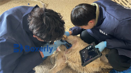
Before egg collection by live B-ultrasound, smL phosphate buffer (PBs) and heparin solution (5 mg/mL) were injected into the collection tube and placed in a 39℃ constant temperature bath. The long needle and catheter were rinsed twice with PBS solution containing heparin and prepared for egg collection. The cow was held in a standing position and epidural anesthesia was performed with smL lidocaine with a concentration of 2%. The operator performed a rectal examination on the cow, cleaned the feces in the rectum, cleaned the vulva, inserted the probe with the egg collection needle into the deep part of the vagina, and placed the probe on the corresponding side of the vaginal ridge according to the position of the ovary to be punctured after reaching the vaginal ridge. The operator held the probe in one hand and pulled the ovary through the rectum with the other hand, pulling the ovary to stick to the probe so that we can clearly see the ovary on the display screen, distinguishing the follicle and CL according to the live B-ultrasound image of egg collection, and adjusting the position of the probe and ovary so that the follicle on the monitor is located on the extension line of the puncture needle straight line, and then the puncture needle is pushed to puncture the follicle. At the same time, the foot switch is used to control the vacuum pump to extract follicular fluid, and the pressure of the vacuum pump is adjusted between 60 and 80 mm Hg. From the monitor, it can be observed that the extracted follicular fluid and follicles begin to shrink and become irregular, and the black image becomes a bright piece until the follicle disappears on the image. The puncture needle is withdrawn, and the second follicle is punctured again to extract follicular fluid until the follicles above Z mm on the ovary on this side are punctured. After the puncture is completed, the probe is adjusted to puncture the follicles on the other side of the ovary in the same way. Finally, after all follicles that meet the conditions have been punctured, the egg collection needle and catheter are rinsed with PBS containing heparin, and the flushing liquid is also poured into the collection tube. The collected follicular fluid is filtered with an embryo collection filter cup, and the oocytes are left in the filter cup and rinsed repeatedly with PBS solution several times. The oocytes in the filter cup are poured into a watch dish, and the eggs are observed under a microscope. The oocytes that meet the requirements are placed in the maturation liquid and matured and cultured in an incubator at 39°C and 5% carbon dioxide saturated humidity.