Ultrasonic diagnosis is a physical examination method that closely combines the physical properties of ultrasound and the acoustic characteristics of the animal's tissue structure. It is divided into B-ultrasound and A-ultrasound. The following introduces the difference between cattle B-ultrasound and veterinary B-ultrasound in cattle operation:
At present, some large-scale dairy farms use cattle B-ultrasound instruments to diagnose early pregnancy in dairy cows. Veterinary B-ultrasound instruments have many advantages in the diagnosis of early pregnancy in dairy cows, such as early diagnosis time, intuitive and reliable cattle B-ultrasound images, early diagnosis, early treatment, and shortened fetal spacing. Reduce some potential losses. During the inspection, the feces in the rectum must be taken out first. Then the probe is inserted into the anus and then slowly sent to the deep part of the rectum. The depth depends on the body size of the dairy cow and the month of pregnancy.
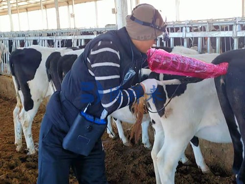
The application of cattle B-ultrasound scanning in dairy cows includes in vitro exploration, vaginal exploration and rectal exploration. Among them, the most suitable for dairy cow examination is rectal exploration. First, restrain the dairy cow to excrete the feces, and then lubricate the probe with lubricant. The operator stands directly behind the cow, holds the probe deep into the rectum, first finds the approximate location of the cow's reproductive organs, and then places the probe close to the top of the reproductive organs for exploration. The depth of the probe varies depending on the size of the cow and the month of pregnancy. The probe chip faces downward, and after finding the uterus, follicles, corpus luteum and other organs, a careful exploration is carried out. During the inspection process, the ambient light should be moderate. If the light is too strong, a cloth should be used to cover the display screen to produce an effective gray shadow effect.
The detection method of veterinary A-ultrasound in Veterinary ultrasound is as follows: The insertion depth of the probe is generally 30-50 cm. It is deeper in multiparous cows than in young cows, and deeper in those with longer gestation periods than in those with shorter gestation periods. Veterinary A-ultrasound detects fetal heart sounds or fetal blood sounds deeper than uterine blood sounds. When randomly detecting, a rectal examination is used for comparison to determine whether the pregnancy is present. The following sounds can be detected in early pregnancy cows: Uterine blood sound (mother's uterine artery blood flow sound) is similar to "ah, ah" and "cicada chirping" sounds, which indicates pregnancy. Its frequency is synchronized with the mother's heart sound, rhythmic, and the sound is vibrating and elongated. If it is similar to "hu-hu-", it is not pregnant; fetal heart sound is like horse hoof sound. It is a rhythmic double beat of "dongdong" and "pudong, pudong". In early pregnancy, it is a single beat or "rustling" sound, which is weak and has no obvious rhythm; fetal blood sound (fetal arterial blood flow sound and umbilical cord artery blood flow sound) is a single beat sound, with a high and sharp tone, rhythmic, and a "beep" sound, which is completely synchronized with the fetal heart sound; fetal movement sound is like a dog barking sound, irregular, and increases with the increase of pregnancy date. The three Doppler signals of uterine blood sound, fetal heart sound and fetal blood sound are the basis for early pregnancy diagnosis. As long as one of these three signals is obtained, pregnancy can be confirmed.