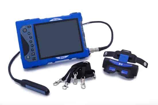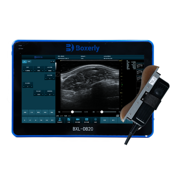
Ultrasound technology plays a crucial role in modern medicine, providing valuable insights into the body’s internal structures. Two common types of ultrasound are Doppler ultrasound and B mode ultrasound. While both serve essential functions in diagnostic imaging, they differ significantly in their applications, methodologies, and the information they provide. This article explores the key differences between Doppler and B mode ultrasound to help you understand their unique roles in healthcare.

## Understanding Ultrasound Basics
Ultrasound imaging utilizes high-frequency sound waves to create images of organs and tissues inside the body. This non-invasive technique is widely used across various medical fields, including obstetrics, cardiology, and emergency medicine.
### What is B Mode Ultrasound?
B mode ultrasound, or brightness mode ultrasound, is one of the most commonly used forms of ultrasound imaging. It generates two-dimensional images by measuring the intensity of reflected sound waves. The resulting grayscale images help healthcare providers visualize the anatomy of organs and tissues, allowing for accurate diagnosis of various conditions.
#### Key Applications of B Mode Ultrasound:
- **Obstetrics:** Monitoring fetal development and assessing placental health.
- **Abdominal Imaging:** Evaluating organs like the liver, kidneys, and gallbladder.
- **Musculoskeletal Imaging:** Diagnosing soft tissue injuries and conditions.
### What is Doppler Ultrasound?
Doppler ultrasound is a specialized technique that assesses blood flow and movement within the body. It measures changes in the frequency of sound waves as they bounce off moving objects, such as red blood cells. This capability allows Doppler ultrasound to visualize and evaluate the velocity and direction of blood flow, making it invaluable in cardiovascular assessments.
#### Key Applications of Doppler Ultrasound:
- **Cardiology:** Evaluating heart conditions, valve function, and blood flow.
- **Obstetrics:** Monitoring fetal heart rate and assessing blood flow in the umbilical cord.
- **Vascular Studies:** Identifying blockages or abnormalities in blood vessels.
## Key Differences Between Doppler and B Mode Ultrasound
### 1. Purpose and Function
- **B Mode Ultrasound:** Primarily focuses on creating static images of anatomical structures. It provides detailed information about the size, shape, and composition of organs and tissues.
- **Doppler Ultrasound:** Specifically designed to assess blood flow and movement. It provides dynamic information about the speed and direction of blood circulation, which is critical for diagnosing vascular conditions.
### 2. Image Representation
- **B Mode Ultrasound:** Produces grayscale images based on the intensity of reflected sound waves. These images allow for a clear view of anatomical structures, helping identify abnormalities.
- **Doppler Ultrasound:** Generates color-coded images that represent the velocity and direction of blood flow. This allows clinicians to visualize how blood moves through the heart and vessels, often overlaying this information on B mode images for a comprehensive assessment.
### 3. Technology and Methodology
- **B Mode Ultrasound:** Utilizes a transducer to emit sound waves, which are reflected back from tissues to create images. The focus is on the density and structure of the tissues.
- **Doppler Ultrasound:** Also uses a transducer but employs the Doppler effect to measure changes in frequency caused by moving blood cells. This allows for real-time visualization of blood flow patterns.
## Conclusion
Doppler and B mode ultrasound are both essential tools in the field of medical imaging, each serving distinct purposes. B mode ultrasound excels in providing detailed anatomical images, while Doppler ultrasound specializes in assessing blood flow and movement. Understanding these differences enables patients and healthcare providers to make informed decisions regarding diagnostic imaging and treatment plans.
### FAQs
**1. Are Doppler and B mode ultrasound safe?**
Yes, both Doppler and B mode ultrasounds are safe and non-invasive procedures that do not use ionizing radiation.
**2. How long do these ultrasound procedures take?**
Typically, both B mode and Doppler ultrasounds take between 15 to 30 minutes, depending on the area being examined.
**3. Do I need special preparation for these ultrasounds?**
Preparation varies depending on the type of ultrasound and the area being examined. Always consult with your healthcare provider for specific instructions.
By understanding the distinctions between Doppler and B mode ultrasound, patients can engage more effectively with their healthcare and better comprehend the diagnostic processes involved in their care.
link: https://www.bxlimage.com/ss/835.html
tags: B Mode Ultrasound Doppler Ultrasound


