An ultrasound probe, also known as a transducer, is a crucial component of ultrasound imaging systems. It is the device responsible for transmitting and receiving the high-frequency sound waves that allow for real-time visualization of internal organs, tissues, and blood flow. In this article, we will explore the function, types, and uses of ultrasound probes, highlighting their importance in medical diagnostics and how they contribute to the accuracy of ultrasound imaging.
")
1. What is an Ultrasound Probe?
An ultrasound probe is a hand-held device that emits sound waves and detects the echoes that bounce back from the body’s tissues. The probe converts these echoes into electrical signals, which are processed by the ultrasound machine to create visual images of the internal structures.
The quality and precision of the ultrasound images depend largely on the type and frequency of the probe used. Different probes are designed for specific applications and areas of the body, and they vary in shape, size, and frequency range.
2. How Does an Ultrasound Probe Work?
The ultrasound probe contains piezoelectric crystals that generate sound waves when an electrical current is applied. These high-frequency sound waves penetrate the body’s tissues and are reflected back to the probe as echoes. The returning echoes are then converted into images displayed on the ultrasound machine’s monitor.
The type of sound waves used by the probe determines the level of detail in the image. Higher-frequency waves provide more detailed images but cannot penetrate as deeply into the body. Lower-frequency waves, on the other hand, can penetrate deeper tissues but may offer less detail.
3. Types of Ultrasound Probes
There are several types of ultrasound probes, each designed for specific diagnostic applications. The choice of probe depends on the body part being examined and the depth of the area to be visualized. Below are some common types of ultrasound probes:
a. Linear Probes
Linear probes emit high-frequency sound waves and are used for superficial structures such as muscles, tendons, blood vessels, and skin. They produce high-resolution images, making them ideal for:
- Vascular ultrasound: Used to assess blood vessels, identify blockages, and evaluate blood flow.
- Musculoskeletal ultrasound: Ideal for imaging soft tissues, such as tendons, ligaments, and muscles.
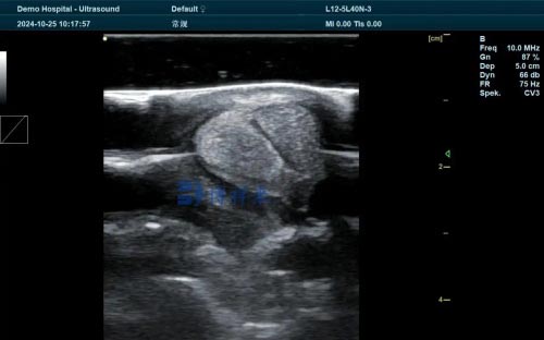
- Thyroid and breast exams: Due to their high resolution, linear probes are effective in detecting abnormalities in small, superficial structures.
b. Curved (Convex) Probes
Curved or convex probes emit lower-frequency waves, allowing them to penetrate deeper into the body. They are commonly used for abdominal, pelvic, and obstetric imaging. Applications include:
- Abdominal ultrasound: Useful for visualizing the liver, kidneys, spleen, and other abdominal organs.
- Obstetric ultrasound: Often used during pregnancy to monitor fetal development and assess the health of the mother’s reproductive organs.
- Pelvic ultrasound: Used to evaluate the uterus, ovaries, and other pelvic structures.
c. Phased Array Probes
Phased array probes are small, compact probes that emit low-frequency waves and are primarily used for cardiac imaging. They allow for imaging between the ribs and provide detailed information on heart function. Their uses include:
- Echocardiography: Phased array probes are essential for diagnosing heart conditions by providing clear images of the heart’s chambers, valves, and blood flow.
- Transcranial Doppler ultrasound: These probes are sometimes used to examine blood flow in the brain.
d. Endocavitary Probes
Endocavitary probes are designed for internal use, offering high-resolution images of internal organs through direct contact with the body’s cavities. These probes are commonly used for:
- Transvaginal ultrasound: Used to visualize the female reproductive system, including the uterus and ovaries, and detect conditions like fibroids, cysts, and early pregnancy.
- Transrectal ultrasound: Frequently used to evaluate the prostate gland in men and assess rectal abnormalities.
e. 3D and 4D Probes
3D and 4D ultrasound probes use advanced technology to create three-dimensional or real-time moving images. These probes are especially useful in obstetric imaging, where they provide detailed images of the fetus, but they also have applications in other areas such as:
- Gynecology: For diagnosing uterine and ovarian abnormalities.
- Cardiology: 3D echocardiography provides detailed images of the heart’s structure and function.
4. Applications of Ultrasound Probes
Ultrasound probes are used in a wide range of medical fields, making them essential tools for diagnostics and treatment planning. Below are some of the key applications:
a. Pregnancy Monitoring
Ultrasound probes are critical in obstetrics, where they help monitor fetal development, detect any complications, and assess the health of the mother’s reproductive organs. They can also be used to determine the position and growth rate of the fetus throughout pregnancy.
b. Cardiac Imaging
Echocardiography probes allow healthcare providers to visualize the heart’s chambers, valves, and blood flow. These probes are invaluable in diagnosing heart conditions, such as heart disease, valve abnormalities, and pericardial effusion.
c. Abdominal Imaging
Curved probes are used to image organs in the abdominal cavity, such as the liver, kidneys, gallbladder, and pancreas. Abdominal ultrasound is often used to detect gallstones, liver disease, kidney stones, and other conditions.
d. Musculoskeletal Imaging
Linear probes are used to assess soft tissue injuries, such as torn ligaments, tendonitis, and muscle strains. They also play a role in guiding injections for pain relief or in performing biopsies.
e. Vascular Studies
Doppler probes are used to measure blood flow through arteries and veins. This helps diagnose conditions like deep vein thrombosis (DVT), blocked arteries, or varicose veins.
5. How to Choose the Right Ultrasound Probe
When selecting an ultrasound probe, several factors need to be considered, including the area of the body being examined, the depth of the tissues, and the level of detail required. Some key considerations include:
- Frequency: Higher frequencies provide better image resolution but are ideal for superficial tissues. Lower frequencies are used for deeper tissues but may sacrifice some image clarity.
- Field of view: The type of probe determines how wide or narrow the viewing area will be, which is important for the specific application.
- Compatibility with ultrasound machines: Ensure the probe is compatible with the ultrasound machine you are using to avoid any technical issues.
6. Maintenance of Ultrasound Probes
Proper maintenance is essential to ensure the longevity and performance of ultrasound probes. Here are some tips:
- Regular Cleaning: After each use, the probe should be cleaned with appropriate disinfectants to prevent infection.
- Careful Handling: Probes are delicate devices and should be handled carefully to avoid damage, especially the crystals inside that generate sound waves.
- Storage: Probes should be stored in protective cases or holders to avoid accidental damage when not in use.
Conclusion
Ultrasound probes are vital components in medical imaging, offering a non-invasive, real-time way to visualize internal structures and diagnose various conditions. From pregnancy monitoring and cardiac health evaluations to musculoskeletal injury assessment, ultrasound probes are used in numerous medical fields. Understanding the different types of probes and their specific applications is essential for healthcare providers to make accurate diagnoses and ensure optimal patient care.
link: https://www.bxlimage.com/ss/842.html
tags: ultrasound probe types of ultrasound probes transducer medical ultrasound linear probe curved probe phased array probe ultrasound transducer