The methods used by veterinary B-ultrasound machines for different objects are also different. So how to use veterinary B-ultrasound machines when testing pigs, sheep and cattle? The following is a detailed introduction: veterinary B-ultrasound machine detection method for sows:
1. Restraint: Sows generally do not need to be restrained, as long as they remain quiet. The posture is best to lie on the side, and it can be crawling, standing or feeding. For individual sows that are difficult to approach, you can use a pig grabber, a mouth rope or a door panel to squeeze into the corner for exploration. Under the conditions of large-scale pig farming, it can be carried out in a restricted feeding pen. When exploring the rectum, the sow needs to stand and restrain.
2. Exploration site: External exploration is generally around the lower abdomen, the upper part of the breast in front of the posterior flank, starting from the posterior and upper part of the last pair of mammary glands (commonly known as milk bags). As pregnancy progresses, the exploration site gradually moves forward and can finally reach the back end of the ribs. Pigs have sparse hair, so there is no need to cut the hair during exploration, but the exploration site must be kept clean. Scrape off dirt and dirt, and apply coupling agent during exploration. Rectum


When exploring inside, it is necessary to ensure that it is fixed. No coupling agent is required. The probe can be moistened with disinfectant.
3. Exploration method: When exploring outside the body, the probe is close to the abdominal wall. When exploring in early pregnancy, the probe is facing the front edge of the pubic bone, the direction of the pelvic entrance, or at a 45-degree angle to the opposite side. The probe is close to the skin and performs fixed-point fan-shaped scans in front and back and up and down. The movement should be slow. The embryo in early pregnancy is very small, and Doppler exploration must be carefully and slowly scanned to detect it. Do not slide the probe on the skin
Quick scan. The A-type instrument should not move too fast when exploring the fetal sac. Sometimes the probe needs to be pressed against the abdominal wall and pressed inward to squeeze the intestinal tube to get closer to the uterus and increase the detection rate. Because before 28-30 days of pregnancy, the uterus usually has not sagged and touched the abdominal wall. After one point exploration is completed, the probe moves forward half to a milk bag to continue exploration until the diagnosis is confirmed.
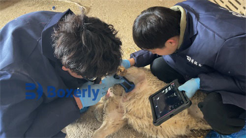
Veterinary B-ultrasound machine detection method for ewes
1. Restraint: Ewes generally take a natural standing posture, with assistants supporting them and keeping quiet, or assistants use their legs to clamp the neck of the ewe for restraint, or use a simple restraint frame for restraint. Side-lying restraint can slightly advance the diagnosis date and improve the accuracy of diagnosis, but it is inconvenient to use in large groups. Type B exploration of early pregnancy can be done in side-lying, supine or standing positions.
2. Exploration site and method: Abdominal wall exploration, in early pregnancy, on both sides of the breast and the hairless area directly in front of the breast, or between the two breasts. In the middle and late stages of pregnancy, it can be performed on the right abdominal wall. There is no need to shear hair in the hairless area, but shearing is required for lateral abdominal wall exploration, and restraint is required for rectal exploration. The exploration method is basically the same as that for pigs. The examiner squats on one side of the sheep body, applies coupling agent locally or on the probe, and then places the probe close to the skin, facing the pelvic inlet, and performs a fixed-point fan-shaped scan. Scanning can be done from the front to the back of the breast, from both sides of the breast to the middle, or from the middle of the breast to both sides. In the early stage of pregnancy, the fetal sac is not large and the embryo is very small, so it needs to be scanned slowly and carefully to be detected. The examiner can also squat behind the buttocks of the goat, hold the probe and extend it from the middle of the two hind legs to the breast for scanning. If the udder of the dairy goat is too large, or the hair on the side of the abdominal wall is too long, which affects the clear view of the exploration site, the assistant can lift the hind leg of the exploration side to expose the exploration site, but there is no need to shear the hair.
Exploration method for predicting the number of fetuses: Ewes do not carry as many fetuses as pigs, and the exploration method is slightly different. The ewe changes to supine position, or semi-supine position, fully exposing the area from the front of the breast to the navel, and can be appropriately sheared. Coupling agent is applied to a larger area, and generally scans from the midline to both sides. D-type instrument exploration determines single or multiple fetuses by detecting the fetal heart beat and the location detected. Do not judge simply by the difference in fetal heart rate, let alone by the frequency of fetal blood sound, as errors are prone to occur. B-type instrument detection, the probe can slide and scan the detection site, and the actual detected embryos are used for judgment. Some people have suggested that the number of fetuses detected in early pregnancy can be used to predict the number of pregnancy, but it is not very reliable.
The probe frequencies are 2.25, 2.5, 3.5, 4.5 and 5.OMHz. 5.0MHz is better in early pregnancy, and 2.25MHz is better in late pregnancy.
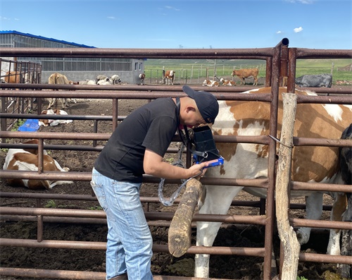
Veterinary B-ultrasound machine detection method for cows
1. Selection of detection site: The uterus of early pregnant cows is located before and after the pelvic entrance. The best site for external detection is the huge udder in front of the pubic bone, the rumen on the left, the intestinal loop on the right, and the thick muscle group on the buttocks, which are far away from the uterus in early pregnancy. Therefore, the best detection site can only be selected in the vagina or rectum close to the uterus. In the middle and late pregnancy, when the uterus sags close to the abdominal wall, the lower abdominal wall can be detected.
2. Exploration method: The cow is restrained in a natural standing position on a cow bed or in a stanchion, just like rectal palpation and rectal grasping and insemination. It can be performed by one person without special restraint. If necessary, an assistant will fix the tail.
Intravaginal exploration: One hand separates the vulva, and the other hand inserts the sterilized long-rod vaginal probe into the vulva, slowly inserting it into the deep vagina and reaching the vault. Type A instrument explores the uterus with fetal fluid. Type D instrument explores the blood flow sound of the uterine artery anastomosis technique at the vault below the cervix on both sides, which is equivalent to 3-5 o'clock and 7-9 o'clock on the clock. Uterine blood sound cannot be detected at the cervix, and generally cannot be detected above the cervix on both sides. Sometimes fetal blood sound or fetal heart sound can be detected below the cervix on both sides. Sanisol solution is often used for probe disinfection. The coupling agent used for intravaginal exploration should be disinfected, but it is generally not necessary. The probes for vaginal exploration are small, and the cows do not feel uncomfortable.
Intrarectal exploration: First stimulate the cow's anus with a long cup rectal probe to induce the cow to defecate, then insert the probe into the anus and then slowly insert it into the deep part of the rectum. When the D-type instrument is used for exploration, the chip surface is first facing upwards, and after passing the lumbar sacral joint, the fan surface is turned to the left or right to explore the middle uterine artery. The depth is generally 30-50cm, and the depth varies depending on the cow's body size and gestation month. The crystal
face is downward and explored to both sides. After 50 days of pregnancy, it is possible to detect fetal blood sound, fetal heart sound or fetal movement sound. When the A-type instrument is used for exploration, the probe chip faces downward, and before and after reaching the pelvic entrance, it is scanned downward and to both sides at a 45-degree angle. When using a D-type or A-type external probe or a ring-type probe for exploration, the hand-worn probe enters the rectum, touches the uterus, and then directly explores the uterine wall. When using a B-type instrument for exploration, a special rectal probe for large animals is held in the hand and brought into the rectum to scan close to the ovaries and uterus. When there is a lot of feces in the rectum, the feces should be discharged first so that the probe can be in close contact with the field wall. When the probe is stained with feces and affects the exploration, it should be removed and cleared. In order to be in close contact with the intestinal wall and protect the probe, it is best to apply coupling agent.
Maintenance methods for veterinary B-ultrasound machines:
1. Life span: According to relevant documents such as design and production, the life span of the product is generally 6 years. The raw materials that constitute the product will gradually age over time. If the product continues to be used after its life span exceeds its life span, it may cause performance degradation and significantly increased failure rate.
2. Check the power cord of the instrument adapter, the probe cable and the waterproof sheath. If damage or breakage is found, it is prohibited to use it and replace it immediately.
Check whether the probe is connected to the host correctly. The power supply of the adapter should be checked frequently. When the power supply voltage exceeds the applicable range specified by the instrument, the adapter shall not be used to power the host or charge the battery. The power adapter is a dedicated power supply with a fully sealed insulation design. It shall not be replaced with other types of adapters at will, and do not try to open the adapter shell to avoid danger.
3. Mainframe maintenance
(1) The use environment should meet the requirements"
(2) When the outer shell needs to be cleaned, it should be wiped with an alcohol cotton ball in the off state.
(3) It is not advisable to open and close the machine frequently.
(4) When not in use for a long time, the instrument should be placed in the packaging box according to the direction indicated on the packaging box and properly stored in the warehouse. The storage environment should meet the requirements.
4. Probe maintenance: The probe is a valuable and vulnerable part. It is strictly forbidden to collide or fall. Medical ultrasonic coupling agent should be used for diagnosis. And the probe shell and jacket should be checked frequently for cracks or damage to avoid immersion in liquid to damage the internal components.
5. Cleaning: When the mainframe and adapter shell need to be cleaned, use a soft dry cloth to wipe the dust on the surface of the body, and then gently wipe it with a 75% medical alcohol cotton ball. The instrument uses a waterproof probe, which can be cleaned by flushing with water or wiping with a wet cloth, but the probe plug cannot be splashed or immersed in water.
6. Use the probe correctly
To extend the life of the probe and obtain the best performance, follow the steps below;
(1) Check the probe cable, socket and acoustic window regularly.
(2) Turn off the probe before connecting or removing it.
(3) Do not drop the probe onto the floor or hard objects. Do not hit the acoustic window of the probe, otherwise it may be damaged.
(4) Do not heat the probe
(5) Do not bend or pull the probe cable, otherwise it may cause the internal connection of the cable to break.
(6) Use coupling agent only on the probe head and clean the probe after use.
(7) After cleaning the probe, carefully check the acoustic window, housing, cable and waterproof sheath of the probe. If cracks or damage are found, do not use it.