The selection principle of the imported backfat analyzer for measuring backfat and eye muscles is: firstly, it should be representative; Secondly, it is easy to detect in vivo; Thirdly, it is less affected by changes in animal position.
The measurement inside or outside the house should be carried out according to the specific situation using appropriate measures. To ensure its natural standing. During the process of moving animals to a stable frame or enclosure, efforts should be made to minimize the occurrence of stress reactions. There are many studies indicating that the optimal location for scanning operations is in the animal enclosure or pen.
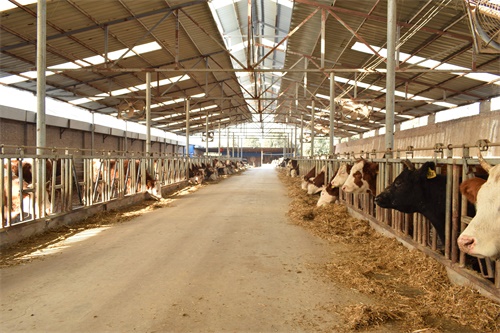
The measurement position of the backfat meter for cattle: the backfat and lumbar muscles are measured about 5cm below the spine behind the twelfth rib. When measuring, place the probe with coupling agent, cut off the hair on the measuring area, and place the probe directly on the body surface. When there is a measuring film, it can also be attached to the probe. Fat thickness measurement is performed between the twelfth and thirteenth ribs, and can be measured horizontally or vertically. When measuring fat thickness horizontally, select an area approximately 3/4 of the lumbar muscle length. The area of the lumbar muscle is measured between the twelfth and thirteenth ribs in the transverse section, and the area of the lumbar muscle is measured in a circle.
Imported backfat analyzer
The measurement position of the backfat meter for pigs: The images of backfat and lumbar muscles can be measured about 5cm below the spine behind the tenth rib.
In order to obtain cross-sectional images of the positive and horizontal axes during measurement, the straight plane of the probe must be perpendicular to the back midline and vertical axis, and must not be slanted. When identifying ultrasound images, first determine the 3-4 strong echo bands generated by the skin interface, adipose tissue, and longest dorsal muscle membrane, and then determine the strong echo images of the muscle membrane around the eye muscle to determine the perimeter of the eye muscle area.
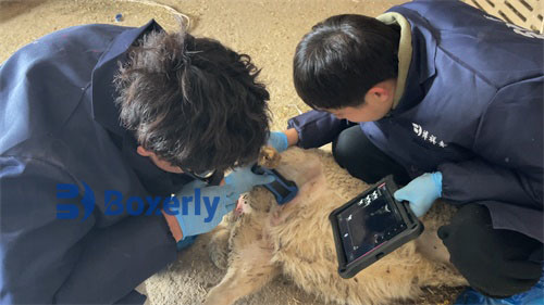
The specific steps for detecting backfat thickness with a portable backfat analyzer are as follows:
Shaving the measurement position - Apply coupling agent (vegetable oil can also be used) to the measurement mold plane and probe plane, so that the measurement mold is adhered to the probe - Apply an appropriate amount of coupling agent to the measurement site to fully immerse it - Place the probe and measurement mold on the measurement position, so that the measurement mold is in close contact with the site, with moderate pressure - Observe and adjust the screen image, adjust the brightness, contrast, and gain appropriately, and try to increase the near-field and decrease the far-field as much as possible. Freeze the image when obtaining the ideal image - Measure the back and waist thickness and eye muscle area, and add explanatory materials (such as measurement time, location, animal number, etc.) - Print or store the image for processing.
Backfat thickness measurement using a backfat analyzer: 3-4 obvious strong echo bands can be seen in ultrasound images, with the first band being the ultrasound reflection at the interface between the measuring membrane and skin, the second band being the fascial reflection between lipids, and the third band being the ocular muscle membrane reflection. According to the screen ruler, the vertical distance of the strong echo point from the l-th shadow band to the 3rd shadow band is the backfat thickness. After determining the starting and ending points during measurement. The millimeter displayed on the screen is the thickness of backfat.
Measurement of eye muscle area using imported backfat analyzer: Due to the strong reflex effect of eye muscle membrane, a clear and approximately elliptical eye muscle contour can be generated in ultrasound images. When measuring, first determine the upper and lower bounds, which are the minor axes of the ellipse, and then determine the boundary points at both ends, which are the major axes of the ellipse. The square centimeter value displayed on the screen at this time is the approximate elliptical area of the eye muscles.